Published online Jun 27, 2013. doi: 10.4240/wjgs.v5.i6.207
Revised: April 16, 2013
Accepted: May 18, 2013
Published online: June 27, 2013
Processing time: 103 Days and 22.6 Hours
Mucocele of the appendix is an uncommon but potentially dangerous pathological entity that presents in a variety of ways. Therefore, optimal surgical therapy is controversial; while some authors adopt a simple appendectomy, others recommend extensive resection, such as right hemicolectomy. We report the case of an 83 years old woman who presented with cystic neoformation in the right iliac fossa that was preoperatively considered an appendiceal mass. We electively performed a laparoscopic resection that histological examination defined as a mucinous cystadenoma. No recurrence was observed in the follow-up period of 9 mo.
Core tip: This report calls the clinician’s attention to the fact that patients presenting with chronic lower right quadrant pain may be diagnosed with appendiceal tumors, particularly elderly patients, as was the case in our report. Since the elderly patient population usually has co-morbid conditions, minimal invasive procedures should be considered after preoperative diagnostic procedures to confirm the absence of advanced tumors.
- Citation: Kaya C, Yazici P, Omeroglu S, Mihmanli M. Laparoscopic appendectomy for appendiceal mucocele in an 83 years old woman. World J Gastrointest Surg 2013; 5(6): 207-209
- URL: https://www.wjgnet.com/1948-9366/full/v5/i6/207.htm
- DOI: https://dx.doi.org/10.4240/wjgs.v5.i6.207
Appendiceal mucocele is an uncommon pathology of the appendix (0.15%-0.6% of all appendectomies) characterized by the accumulation of mucoid material in the lumen[1,2]. Etiopathogenesis can be inflammatory or neoplastic. Dissemination of neoplastic cells and mucoid material in the abdominal cavity caused by appendiceal perforation clinically results in pseudomyxoma peritonei. Therapy is fundamentally surgical and several options have been reported, ranging from simple appendectomy to right hemicolectomy. We report here a case of an 83 years old woman with one month history of right lower abdominal pain caused by an appendiceal mass that was laparoscopically resected.
An 83 years old woman presented with abdominal distension and discomfort in the right lower quadrant of one month duration. In her medical history, she had hypertension for 10 years and rheumatoid arthritis for 2 years. She was taking acetyl salicylic acid and a nonsteroidal anti-inflammatory. She was an active smoker with a 50 pack-year smoking history and was operated on for a perianal fistula 20 years previously. On physical examination, she was afebrile and hemodynamically stable. The abdominal examination was normal except for focal tenderness over McBurney’s point without rebound tenderness on palpation and a mass lesion over the right lower quadrant. Ultrasound examination was performed and a cystic mass in the right iliac fossa of about 4 cm in size was reported.
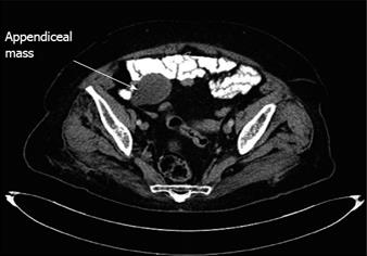
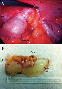
Laboratory analysis was unremarkable. Axial computed tomography (CT) scanning revealed a 4 cm × 3.5 cm × 3 cm, blind ending, tubular, fluid-filled structure that appeared to arise from the cecum, consistent with mucocele of the appendix (Figure 1). Colonoscopy showed evidence of an appendiceal mass covered by cecal mucosa; no other pathology was detected. Intra-operative observation revealed a smooth, cystic tumor of the appendix (Figure 2A). There was no ascites, metastatic peritoneal nodules or ovarian pathology as evidence of malignancy. Laparoscopic excision of the unruptured appendiceal mucocele with the mesoappendix was performed (Figure 2B). We used LigaSure™ for the appendiceal excision but other choices are available, such as simple knotting and cut or stapler use.
Histopathological examination showed low grade epithelial dysplasia, a feature diagnostic of a mucinous cystadenoma, measuring 3.5 cm at its greater diameter. No lymph node involvement was discovered in the appendiceal mesentery. The patient was discharged on postoperative day one and recovered uneventfully. The patient remained well and symptom-free during the follow-up period of 9 mo.
Appendiceal mucocele does not have a typical clinical picture. More than two-thirds of patients have their appendiceal mucocele removed on incidental finding[3]. Clinically, it can remain asymptomatic or manifest with acute or chronic abdominal pain which is the most common clinical finding, as was the case in our patient. In an elderly patient, undoubtedly, malignancy should be considered as a differential diagnosis and preoperative diagnostic methods are needed to avoid inappropriate treatment. Also, risk of perforation of the appendix is significantly higher in elderly patients.
Laparoscopic appendectomy is becoming popular because of its advantages, not only for acute appendicitis but also for perforated appendicitis and even suspected malignant lesions. The optimal treatment modality for appendiceal mucocele is still controversial. Surgical management of this entity differs primarily depending on the characteristics of the mass (location and size) and clinical presentation, whereas the approach used (open or minimally invasive techniques) depends partly upon the preference and experience of the surgeon. Although some authors still recommend open procedures for appendiceal masses[4], particularly for those with possible malignancy, laparoscopic techniques have been described to minimize the risk of seeding tumor implants during laparoscopic manipulation[5]. In our case, preoperative imaging studies were not suspicious for malignancy. CT revealed no involvement of the appendiceal base, mesoappendix or local lymph nodes. We ensured isolation of the appendix, wrapping gauze around the appendiceal structures during laparoscopic resection and then putting it into an endobag to avoid any contamination of the abdominal cavity in case of perforation. Fortunately, histopathological examination revealed a low grade appendiceal mucinous neoplasm confined to the appendix and no further therapy was required. In addition to the diagnostic advantages of laparoscopy, excision of the mesoappendix with the main cystic appendiceal tumor helps assess stage of disease.
In the era of minimally invasive procedures, laparoscopic appendectomy for mucocele should be considered as the primary choice, particularly in elderly patients in the absence of preoperative confirmation of locally advanced malignancy of the appendix. The laparoscopic approach allows diagnostic evaluation and appendectomy to be performed and confers advantages of minimal invasive surgery as well as a short hospital stay and decreased recovery period, particularly in patients with co-morbid conditions.
P- Reviewers Meijerink W, Rousei S S- Editor Zhai HH L- Editor Roemmele A E- Editor Lu YJ
| 1. | Marudanayagam R, Williams GT, Rees BI. Review of the pathological results of 2660 appendicectomy specimens. J Gastroenterol. 2006;41:745-749. [RCA] [PubMed] [DOI] [Full Text] [Cited by in Crossref: 157] [Cited by in RCA: 158] [Article Influence: 8.3] [Reference Citation Analysis (0)] |
| 2. | Rangarajan M, Palanivelu C, Kavalakat AJ, Parthasarathi R. Laparoscopic appendectomy for mucocele of the appendix: Report of 8 cases. Indian J Gastroenterol. 2006;25:256-257. [PubMed] |
| 3. | Stocchi L, Wolff BG, Larson DR, Harrington JR. Surgical treatment of appendiceal mucocele. Arch Surg. 2003;138:585-589; discussion 585-589. [RCA] [PubMed] [DOI] [Full Text] [Cited by in Crossref: 107] [Cited by in RCA: 111] [Article Influence: 5.0] [Reference Citation Analysis (0)] |
| 4. | Sturniolo G, Barbuscia M, Taranto F, Tonante A, Paparo D, Romeo G, Nucera D, Lentini M. Mucocele of the appendix. Two case reports. G Chir. 2011;32:487-490. [PubMed] |
| 5. | Chiu CC, Wei PL, Huang MT, Wang W, Chen TC, Lee WJ. Laparoscopic resection of appendiceal mucinous cystadenoma. J Laparoendosc Adv Surg Tech A. 2005;15:325-328. [RCA] [PubMed] [DOI] [Full Text] [Cited by in Crossref: 22] [Cited by in RCA: 22] [Article Influence: 1.1] [Reference Citation Analysis (0)] |


