Published online Dec 27, 2013. doi: 10.4240/wjgs.v5.i12.329
Revised: November 25, 2013
Accepted: December 12, 2013
Published online: December 27, 2013
Processing time: 71 Days and 12.8 Hours
Free intraperitoneal air is thought to be pathognomonic for perforation of a hollow viscus. Here, we present a patient with pain in the upper left quadrant, a mild fever and leukocytosis. Free air was suggested under the left diaphragm but during the explorative laparotomy no signs of gastric or diverticular perforation were seen. Further exploration and revision of the computed tomography revealed a perforated splenic abscess. Splenic abscesses are a rare clinical entity. Presenting symptoms are often non-specific and include upper abdominal pain, recurrent or persistent fever, nausea and vomiting, splenomegaly, leukocytosis and left lower chest abnormalities. Predisposing conditions can be very divergent and include depressed immunosuppressed state, metastatic or contiguous infection, splenic infarction and trauma. Splenic abscess should therefore be considered in a patient with fever, left upper abdominal pain and leukocytosis. Moreover, our case shows that splenic abscess can present in an exceptional way without clear underlying aetiology and should even be considered in the presence of free abdominal air.
Core tip: Free intraperitoneal air is thought to be pathognomonic for perforation of a hollow viscus. Here, we present a patient with pain in the upper left quadrant, a mild fever and leukocytosis. Free air was suggested under the left diaphragm but during the explorative laparotomy no signs of gastric or diverticular perforation were seen. Further exploration and revision of the computed tomography revealed a perforated splenic abscess. Splenic abscesses are a rare clinical entity. Our case shows that splenic abscess can present in an exceptional way without clear underlying aetiology and should even be considered in the presence of free abdominal air.
- Citation: Nunspeet LV, Eddes EH, Noo ME. Uncommon cause of pneumoperitoneum. World J Gastrointest Surg 2013; 5(12): 329-331
- URL: https://www.wjgnet.com/1948-9366/full/v5/i12/329.htm
- DOI: https://dx.doi.org/10.4240/wjgs.v5.i12.329
Splenic abscess is a rare condition with a reported frequency in autopsy series between 0.1% to 0.7%[1-3]. Presenting symptoms include upper abdominal pain, recurrent or persistent fever, nausea and vomiting, splenomegaly, leukocytosis and left lower chest abnormalities[4,5]. Diagnosis of a splenic abscess is confirmed on ultrasound or computed tomography (CT)-imaging of the abdomen. Splenectomy has been the gold standard treatment for splenic abscess, however more recent percutaneous drainage is also suggested to be safe and effective[1,6,7]. While gas formation in splenic abscess has been described, few have reported pneumoperitoneum as presenting symptom of a ruptured splenic abscess[8-11].
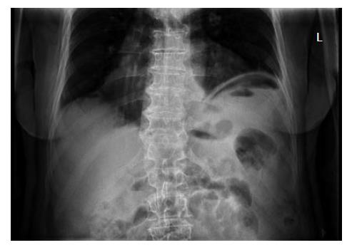
A 78-year-old man presented to our Emergency Room with acute abdominal pain located in the upper left quadrant. The pain had presented in the middle of the night, waking the patient. No nausea nor vomiting had occurred, but he was experiencing an urge to move. His clinical record mentioned a mild mitralis valve insufficiency, atypical rheumatic complains and diverticulosis. He did not use immunosuppressive medication. Clinical examination reported a painful man with a mild fever and raised pulse. The abdomen was bloated, showed little peristaltic sounds, while percussion of the liver was normal and neither liver nor spleen were palpable. Laboratory findings showed leukocytosis and a raised CRP. On the standing X-ray of the thorax a strong suspicion of free air was suggested under the diaphragm, which was confirmed with an X-ray of the abdomen in left lateral position (Figure 1).
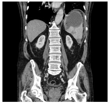
Additional CT showed free air in the upper abdomen with some abdominal fluid left paracolic and in the small pelvis, some left pleural effusion, thickening of the gastric wall and a cyst in the spleen (Figure 2). Therefore, a gastric perforation was suggested. There was no sign of diverticular infiltration or perforation.
After intravenous antibiotics were started on the ER, an explorative laparotomy was performed. No signs of gastric or diverticular perforation were seen. Re-evaluation of the CT in the operation room was performed and the suggestion of an abscess rather than a cyst in the spleen was introduced (Figure 2). Further exploration of the flexura lienalis was performed and pus was evacuated from the upper left quadrant. A ruptured splenic abscess was found and a splenectomy was performed. Cultures remained negative for any grow of bacteria. Pathology report of the spleen revealed an inflammation with abscess and necrosis without micro-organisms or signs of neoplasia. Post-splenectomy vaccinations were prescribed and the patient was discharged 2 wk after admission. Two months after surgery he was in a good clinical condition.
Diagnosis of splenic abscess is often not considered due to its rarity and the presence of predisposing conditions which obscure its clinical presentation[6]. Thereby, the aetiology of splenic abscesses is diverse. Three etiological causes of splenic abscesses have been proposed by Kuttner: trauma with secondary infection; per continuitatem; and haematogenous spread[12]. Development by continuitatem has been described in perforated gastric ulcer, perinephric abscess, septic abortion, appendicitis with perforation and in case of concomitant colon carcinoma[1,3,13,14]. Colon carcinoma are also important precursors in the small number of cases in which metastasis of the spleen were secondary infected[15]. Other haematological spread can be caused by retropharyngeal abscess, otitis media, tonsillectomy, infective endocarditis and phlebitis of the calf[3,5,16].
The most common organisms found on bacteriological examination are Gram Negative Bacillus (Klebsiella Pneumoniae, Escherichia Coli) and Gram Positive Coccus (Staphylococcus Aureus), although a great variety of pathogens have been described[4,17,18].
All studies on this subject stress the strong correlation between splenic abscess and predisposing factors. Direct trauma, infarction or ischemia of the spleen predispose to secondary infection. Especially immunosuppressive state seems to play a great role in the development and rising incidence of splenic abscesses[19]. Furthermore, intravenous drug abuse, human immunodeficiency virus, diabetes mellitus, tuberculosis and neoplasia seem to be contributing diseases[4,8,15,20].
Review of the literature shows only a few cases in which a splenic abscess presented with a pneumoperitoneum[8-11]. In some of these cases the aetiology is clear, but all needed an explorative laparotomy to clarify the diagnosis.
In our case, due to the free abdominal air we expected to find a gastric perforation. The splenic abscess was detected during the explorative laparotomy and only in retrospection the CT-images were interpreted accordingly. Postoperative evaluation revealed no aetiological cause of the splenic abscess. The patient did have diverticulosis, but on operative inspection no inflammation was present. Pathology report of the spleen revealed an inflammation with abscess and necrosis without micro-organisms or signs of neoplasia. Futhermore, blood cultures remained negative in our case. This appears to be the case in approximately 30% of patients with a splenic abscess[4,5]. In conclusion, splenic abscess should be considered in a patient with fever, left upper abdominal pain, and leukocytosis[7]. Moreover, our case shows that splenic abscess can present in an exceptional way without clear underlying aetiology and should even be considered in the presence of free abdominal air.
The presenting symptoms include acute abdominal pain located in the upper left quadrant with and an urge to move.
The patient had a mild fever and raised pulse, a bloated abdomen which showed little peristaltic sounds.
Based on these findings an extensive differential diagnosis of intra-abdominal pathology arose.
Laboratory findings showed a leukocytosis and raised CRP. On the standing X-ray of the thorax free air was suggested and a strong suspicion of perforation of a hollow viscus arose.
Additional computed tomography showed free air in the upper abdomen with some abdominal fluid left paracolic and in the small pelvis, thickening of the gastric wall and a cyst in the spleen.
Review of the literature shows only a few cases in which a splenic abscess presented with a pneumoperitoneum. In some of these cases the aetiology is clear, but all needed an explorative laparotomy to clarify the diagnosis.
After intravenous antibiotics were started, an explorative laparotomy was performed and a ruptured splenic abscess was treated by a splenectomy.
While gas formation in splenic abscesses has been described, few have reported pneumoperitoneum as presenting symptom of a ruptured splenic abscess.
Therefore, splenic abscess should be considered in a patient with fever, left upper abdominal pain and leukocytosis, even in the presence of free abdominal air.
This is a very interesting case report.
P- Reviewers: Al-Mufarrej FMI, Jiang X, Pavlidis TE S- Editor: Cui XM L- Editor: A E- Editor: Wang CH
| 1. | Sreekar H, Saraf V, Pangi AC, Sreeharsha H, Reddy R, Kamat G. A retrospective study of 75 cases of splenic abscess. Indian J Surg. 2011;73:398-402. [RCA] [PubMed] [DOI] [Full Text] [Cited by in Crossref: 13] [Cited by in RCA: 16] [Article Influence: 1.1] [Reference Citation Analysis (0)] |
| 2. | Carbonell AM, Kercher KW, Matthews BD, Joels CS, Sing RF, Heniford BT. Laparoscopic splenectomy for splenic abscess. Surg Laparosc Endosc Percutan Tech. 2004;14:289-291. [RCA] [PubMed] [DOI] [Full Text] [Cited by in Crossref: 33] [Cited by in RCA: 30] [Article Influence: 1.5] [Reference Citation Analysis (0)] |
| 3. | Knauer QF, Abrams JS. Generalized peritonitis due to a ruptured splenic abscess. Am J Surg. 1966;112:923-926. [RCA] [PubMed] [DOI] [Full Text] [Cited by in Crossref: 5] [Cited by in RCA: 5] [Article Influence: 0.1] [Reference Citation Analysis (0)] |
| 4. | Chang KC, Chuah SK, Changchien CS, Tsai TL, Lu SN, Chiu YC, Chen YS, Wang CC, Lin JW, Lee CM. Clinical characteristics and prognostic factors of splenic abscess: a review of 67 cases in a single medical center of Taiwan. World J Gastroenterol. 2006;12:460-464. [PubMed] |
| 5. | Lee WS, Choi ST, Kim KK. Splenic abscess: a single institution study and review of the literature. Yonsei Med J. 2011;52:288-292. [RCA] [PubMed] [DOI] [Full Text] [Full Text (PDF)] [Cited by in Crossref: 60] [Cited by in RCA: 63] [Article Influence: 4.5] [Reference Citation Analysis (0)] |
| 6. | Fotiadis C, Lavranos G, Patapis P, Karatzas G. Abscesses of the spleen: report of three cases. World J Gastroenterol. 2008;14:3088-3091. [RCA] [PubMed] [DOI] [Full Text] [Full Text (PDF)] [Cited by in CrossRef: 35] [Cited by in RCA: 28] [Article Influence: 1.6] [Reference Citation Analysis (0)] |
| 7. | Conzo G, Docimo G, Palazzo A, Della Pietra C, Stanzione F, Sciascia V, Santini L. The role of percutaneous US-guided drainage in the treatment of splenic abscess. Case report and review of the literature. Ann Ital Chir. 2012;83:433-436. [PubMed] |
| 8. | Ishigami K, Decker GT, Bolton-Smith JA, Samuel I, Wilson SR, Brown BP. Ruptured splenic abscess: a cause of pneumoperitoneum in a patient with AIDS. Emerg Radiol. 2003;10:163-165. [RCA] [PubMed] [DOI] [Full Text] [Cited by in Crossref: 9] [Cited by in RCA: 9] [Article Influence: 0.4] [Reference Citation Analysis (0)] |
| 9. | Rege SA, Philip U, Quentin N, Deolekar S, Rohandia O. Ruptured splenic abscess presenting as pneumoperitoneum. Indian J Gastroenterol. 2001;20:246-247. [PubMed] |
| 10. | Puhakka KB, Boljanovic S. Ruptured splenic abscess as cause of pneumoperitoneum. Rofo. 1997;166:273-274. [RCA] [PubMed] [DOI] [Full Text] [Cited by in Crossref: 1] [Cited by in RCA: 2] [Article Influence: 0.1] [Reference Citation Analysis (0)] |
| 11. | Braat MN, Hueting WE, Hazebroek EJ. Pneumoperitoneum secondary to a ruptured splenic abscess. Intern Emerg Med. 2009;4:349-351. [RCA] [PubMed] [DOI] [Full Text] [Cited by in Crossref: 5] [Cited by in RCA: 3] [Article Influence: 0.2] [Reference Citation Analysis (0)] |
| 12. | van de Wielt W, van Dongen R. Splenic abscess as a complication of salmonella infection. Ned Tijdschr Geneeskd. 1964;108:992-994. [PubMed] |
| 13. | Giacobbe A, Facciorusso D, Modola G, Caturelli E, Caruso N, Perri F, Tardio B, Bisceglia M, Andriulli A. Splenic abscess secondary to penetrating gastric ulcer. Minerva Gastroenterol Dietol. 1998;44:111-115. [PubMed] |
| 14. | Stewart IE, Borland C. Case report: perinephric-splenic fistula--a complication of percutaneous perinephric abscess drainage. Br J Radiol. 1994;67:894-896. [RCA] [PubMed] [DOI] [Full Text] [Cited by in Crossref: 1] [Cited by in RCA: 1] [Article Influence: 0.0] [Reference Citation Analysis (0)] |
| 15. | Pisanu A, Ravarino A, Nieddu R, Uccheddu A. Synchronous isolated splenic metastasis from colon carcinoma and concomitant splenic abscess: a case report and review of the literature. World J Gastroenterol. 2007;13:5516-5520. [PubMed] |
| 16. | Robinson SL, Saxe JM, Lucas CE, Arbulu A, Ledgerwood AM, Lucas WF. Splenic abscess associated with endocarditis. Surgery. 1992;112:781-786; discussion 786-787. [PubMed] |
| 17. | Brook I, Frazier EH. Microbiology of liver and spleen abscesses. J Med Microbiol. 1998;47:1075-1080. [RCA] [PubMed] [DOI] [Full Text] [Cited by in Crossref: 128] [Cited by in RCA: 105] [Article Influence: 3.9] [Reference Citation Analysis (0)] |
| 18. | Nelken N, Ignatius J, Skinner M, Christensen N. Changing clinical spectrum of splenic abscess. A multicenter study and review of the literature. Am J Surg. 1987;154:27-34. [RCA] [PubMed] [DOI] [Full Text] [Cited by in Crossref: 164] [Cited by in RCA: 136] [Article Influence: 3.6] [Reference Citation Analysis (0)] |
| 20. | Phillips GS, Radosevich MD, Lipsett PA. Splenic abscess: another look at an old disease. Arch Surg. 1997;132:1331-1335; discussion 1335-1336. [RCA] [PubMed] [DOI] [Full Text] [Cited by in Crossref: 60] [Cited by in RCA: 63] [Article Influence: 2.3] [Reference Citation Analysis (0)] |


