Published online Apr 27, 2011. doi: 10.4240/wjgs.v3.i4.54
Revised: March 24, 2011
Accepted: March 30, 2011
Published online: April 27, 2011
A bezoar is an intraluminal mass formed by the accumulation of undigested material in the gastrointestinal tract. A trichobezoar is a bezoar made up of hair and is a rare cause of bowel obstruction of the proximal gastrointestinal tract. They are seen mostly in young women with trichotillomania and trichotillophagia and symptoms include epigastric pain, nausea, loss of appetite and bowel or gastric outlet obstruction. We herein describe a case of a trichobezoar that presented as a gastric outlet obstruction and was subsequently successfully removed via a laparotomy.
- Citation: Gaujoux S, Bach G, Au J, Godiris-Petit G, Munoz-Bongrand N, Cattan P, Sarfati E. Trichobezoar: A rare cause of bowel obstruction. World J Gastrointest Surg 2011; 3(4): 54-55
- URL: https://www.wjgnet.com/1948-9366/full/v3/i4/54.htm
- DOI: https://dx.doi.org/10.4240/wjgs.v3.i4.54
A bezoar is an intraluminal mass formed by the accumulation of undigested material in the gastrointestinal tract. It can be composed of vegetable fibers (phytobezoar), medication (pharmacobezoar), undigested milk (lactobezoar) or foreign material such as hair (trichobezoar).
Trichobezoars are seen mostly in young women with trichotillomania and trichotillophagia and symptoms include epigastric pain, nausea, loss of appetite and bowel or gastric outlet obstruction. Trichobezoars are a rare cause of bowel obstruction of the proximal gastrointestinal tract. We herein describe a case of a trichobezoar that presented as a gastric outlet obstruction and was successfully removed via a laparotomy.
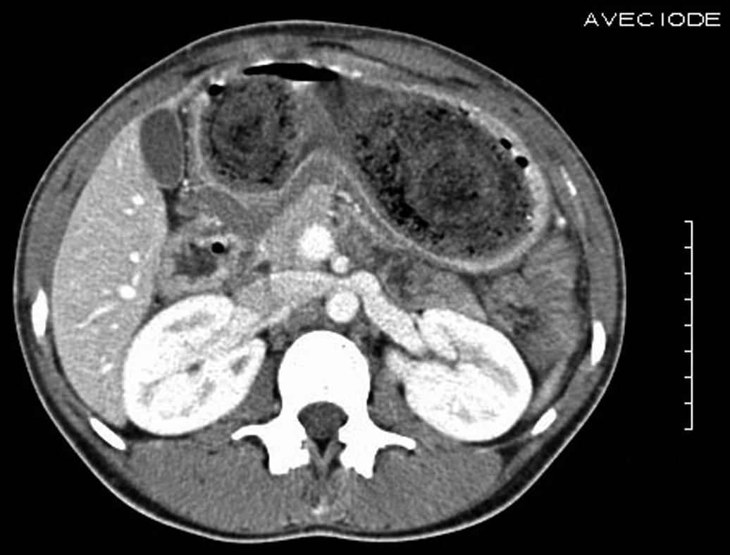
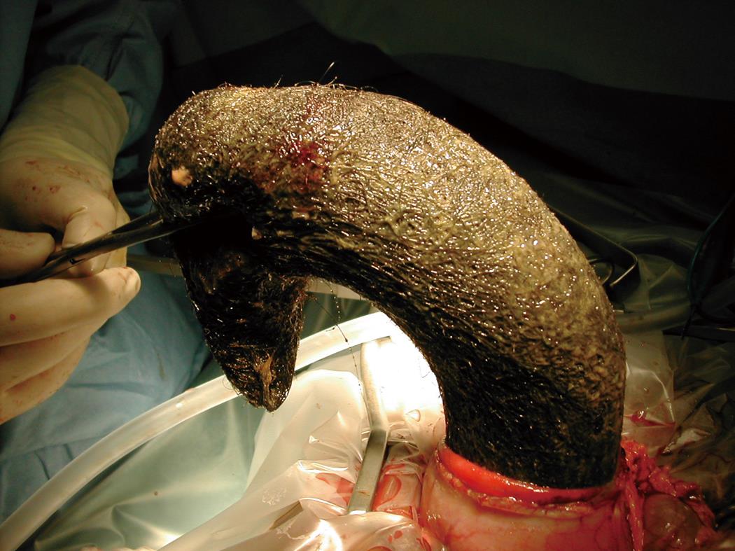
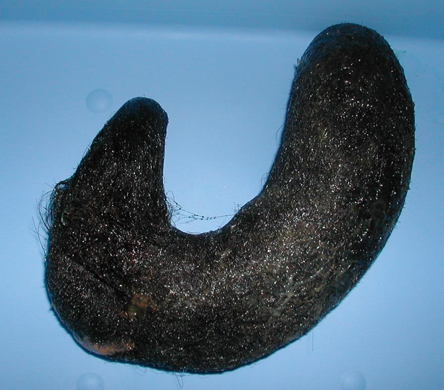
A 19-year-old woman with no past medical history presented to the hospital with bowel obstruction. Clinical examination revealed a painful epigastric mass which on computed tomography (CT) scan was heterogenous, hypodense, non-enhancing and inside the stomach (Figure 1). Due to its bulky size, the decision was made to proceed with a laparotomy. During the surgery, there was no evidence of perforation or ischemia of the stomach but the stomach was full with a soft mass. A horizontal gastrotomy was made at the fundus and a trichobezoar that had assumed the shape of the stomach was discovered and successfully extracted (Figure 2 and Figure 3). The trichobezoar weighed 1250 g and was 21 cm long. The patient’s postoperative course was uneventful and the patient later admitted to a history of trichotillomania and trichotillophagia, for which she then received counseling.
Trichobezoars, undigested accumulations of hair in the gastrointestinal tract, are the most common type of bezoars, commonly seen in patients under 30 years of age[1]. In 90% of cases, the patients are women with long hair and emotional or psychiatric disorders. Clinical manifestations are non-specific - abdominal pain, nausea, constipation - but trichobezoars can lead to serious complications such as bowel obstruction, hemorrhage or perforation[2,3].
In the majority of cases, the diagnosis is based on review of the past medical history and a CT scan of the abdomen with oral contrast[4]. Trichobezoars are generally oval, intraluminal, well delineated masses that are heterogenous with densities of both air and soft tissue. They are outlined by ingested contrast but are themselves non-enhancing. It is easier to make the diagnosis if using a wide window above 400 UH on CT to see the bubbles of air trapped within the bezoar. Other information offered by the CT scan includes the number of bezoars, their location, which are mostly intragastric with occasional extension to the small bowel (Rapunzel syndrome), and any complications. The treatment of choice is surgical removal and the size of trichobezoars often necessitates a laparotomy for removal. Endoscopic fragmentation may be attempted but often fails[1].
Peer reviewer: Thomas J Miner, MD, FACS, Department of Surgery, Rhode Island Hospital, 593 Eddy Street - APC 439, Providence, RI 02903, United States
S- Editor Wang JL L- Editor Roemmele A E- Editor Zheng XM
| 1. | Andrus CH, Ponsky JL. Bezoars: classification, pathophysiology, and treatment. Am J Gastroenterol. 1988;83:476-478. |
| 2. | Krausz MM, Moriel EZ, Ayalon A, Pode D, Durst AL. Surgical aspects of gastrointestinal persimmon phytobezoar treatment. Am J Surg. 1986;152:526-530. |
| 3. | Escamilla C, Robles-Campos R, Parrilla-Paricio P, Lujan-Mompean J, Liron-Ruiz R, Torralba-Martinez JA. Intestinal obstruction and bezoars. J Am Coll Surg. 1994;179:285-288. |


