INTRODUCTION
Minimally invasive surgery represents a significant shift in surgical approach from the conventional open surgery methods, offering patient benefits including reduced morbidity, shorter hospital stays, and better cosmetic results. The safety and efficacy of laparoscopic techniques has been demonstrated in several abdominal surgeries[1-4]. Adequate access to target organs in the abdominal space, either for diagnostic or therapeutic purposes, is critical for surgical intervention. In contrast to traditional laparotomy, laparoscopic techniques employ strategic arrangement of limited ports for abdominal cavity access depending on the surgical procedure. The notion that safety and efficacy of current laparoscopic techniques can be further enhanced by reducing the number of laparoscopic port incisions[5,6] has resulted in the renewed interest in single incision laparoscopic surgery for several surgical procedures such as cholecystectomy, appendectomy and nephrectomy[7-10]. However, outcomes of single incision surgery are still being evaluated.
Consistent with a push towards further reducing surgical invasiveness, natural orifice surgery is a novel surgical approach. Natural orifice translumenal endoscopic surgery (NOTES) involves the use of natural orifices to gain access to the abdominal cavity[11]. This effectively eliminates the need for any external incisions for visceral access, and thus NOTES represents the least invasive surgical technique. Successful application of NOTES technology has been demonstrated in several pre-clinical studies using either a transgastric, transvesical or transcolonic approach[12-14]. A NOTES approach has also been used for various abdominal surgical procedures such as organ resection[15], gastrojejunostomy and gastrojejunal anastomosis[16,17], and oopherectomy and tubectomy[18]. Recently, NOTES has shown promise in clinical studies of cholecystectomy and appendectomy[19,20]. Despite several studies, NOTES experience remains largely experimental with limited clinical studies.
FLEXIBLE ENDOSCOPY PLATFORM FOR NOTES
Although several experimental studies have clearly demonstrated the feasibility and successful application of NOTES[12-18], inherently the approach appears to be surgically challenging. NOTES approach, in principle, shows an amalgamation of laparoscopic minimally invasive surgery techniques with endoscopic technology. Recent advances in fiberoptics and endoscopic technology allow for enhanced imaging and manipulation of visceral organs. A typical NOTES procedure incorporates a flexible endoscopy platform to obtain access to the peritoneal cavity. Current flexible video-endoscopes are specifically designed to traverse through hollow intraluminal structures and provide image-guided navigation through body cavities. With the help of a trans-visceral surgical incision, an entry into the abdominal space is accomplished. Subsequently, the accessory channels of the flexible endoscope are utilized to insert devices for manipulation of the target abdominal organ. Upon completion of the surgical tasks, the visceral incision is repaired and the scope manually withdrawn through the natural orifice[11]. However, several questions regarding the technique still remain unanswered. The optimal visceral site for access to the peritoneal cavity remains unclear. Consequently, there is lack of a standardized approach for creating this trans-visceral opening. Additionally, lack of adequate closure techniques for the NOTES visceral incision site has resulted in limited applications of this surgical endoscopic technique[11].
NOTES INSTRUMENTATION
Despite the advances in NOTES technology, this novel surgical approach exhibits a unique instrumentation paradox. Progressive propagation of instruments first through the hollow visceral lumen followed by maneuverability to different parts of the abdomen through a visceral incision requires the design and development of endoscopic entry tools that are flexible in their entirety. However, once within the anatomic space, the same instruments are required to provide a stable platform for organ and tissue manipulation. As a result endoscope fixation and stiffening may be optimal for such tasks[21]. In addition, there is inadequate visualization of the surgical environment during the NOTES procedure. The prospect of working through a small orifice and within a closed lumen while also performing tasks that require precision is surgically challenging. The problem is further compounded by two-dimensional imaging of the operating field and limited intuitive knowledge of the exact orientation of the scope within the lumen. Furthermore, organ and tissue manipulation require precise articulation and triangulation capabilities of surgical tools. These capabilities, in addition, should not be restricted in any fashion by the movement of the endoscope. However, current flexible video-endoscopes offer minimal triangulation and hence impair surgical manipulation. Current flexible endoscopy platforms allow for accessory instrumentation through the endoscope channels that are in the same geometric plane as the endoscope. This single-planar instrumentation further restricts the ability to apply off-axis forces for organ manipulation[11]. Thus, inadequate visualization and lack of triangulation with existing instruments limits surgical dexterity. Therefore, there is a need for new and improved surgical tools and instrumentation for NOTES.
FLEXIBLE ROBOTICS PLATFORM FOR NOTES
Limited visualization and surgical dexterity constraints were similarly seen during the early development of laparoscopic surgery. Robotic technology significantly influenced laparoscopic surgery and alleviated some of these constraints of traditional laparoscopic surgery. The commercially available da Vinci Surgical System (Intuitive Surgical Inc., Sunnyvale, CA) incorporated features such as articulating end effectors and increased degrees of freedom that significantly increased surgical dexterity during laparoscopic surgery. In addition, stereoscopic three-dimensional vision and tremor abolition enabled sophisticated visual feedback during surgical tasks[22]. These enhancements and the development of robotic instruments and devices further improved tissue and target organ manipulation during laparoscopic surgical tasks.
Similarly, the application of robotic technology to existing NOTES technique can revolutionize natural orifice surgery. As with laparoscopic surgery, robotics can help address some of the classical constraints of the flexible endoscopy platform used during NOTES. Better visualization through robotic stereoscopic visualization and significant improvement in surgical dexterity by enhancing degrees of freedom for complex surgical tasks can be accomplished with a flexible robotics platform[23]. Another critical area that could benefit from a flexible robotics platform is the mechanical control of endoscope movement within the tubular lumen. Existing flexible video-endoscopes used in the NOTES approach allow for manual control of endoscope motion. This is best suited for traversing less complex, tubular, hollow structures during endoscopy procedures and is certainly suboptimal for complex, small anatomic spaces. Manual control of existing endoscopic technology is also not suitable for navigation through a three-dimensional, complex abdominal cavity that requires several maneuvers and fine control of the endoscope tip. Constant positioning and re-positioning of the manual endoscope in several desired locations of the abdominal cavity remains troublesome. As a result, current manipulation and steering technologies are crude and do not offer the surgical precision required for complex surgical tasks during NOTES. Thus, development of a flexible robotics platform capable of providing better visualization, precision maneuverability in large cavities, fine motor control of endoscope distal tip during complex tasks and enhanced surgical dexterity is necessary[24].
MINIATURE IN VIVO SURGICAL ROBOTS FOR NOTES
Although a flexible robotics platform can offer significant advantages over the current platform, there are specific limitations that may hamper this technology. Current surgical robotics is largely an exoluminal technology that is further constrained by the large size of the robot. Despite the advantages of increased degrees of freedom, robotics technology still remains, to some extent, constrained by the fulcrum effect at the abdominal wall incision. Animal studies in a porcine model of NOTES nephrectomy have noted some of these limitations. A transvaginal NOTES approach for porcine pyeloplasty and nephrectomy with the da Vinci robotic platform has demonstrated frequent collisions of robotic arms and raised the issue of appropriateness of the da Vinci surgical platform for NOTES[25]. These results suggest that in the present form, any application of surgical robotics for NOTES seems inappropriate because the available technology is too large for the natural orifice and does not confirm to the geometry of the lumen. Miniaturization of the robotic technology can significantly enhance the degrees of freedom in either a laparoscopic or NOTES procedure[21]. A fundamentally different and alternative approach to access the abdominal viscera for either a laparoscopic or NOTES procedure is the use of small robotic devices that can be introduced in an intracorporeal fashion. These robots can be inserted in the abdominal cavity and are thus not constrained by the abdominal wall incision. Additionally multiple, independent, miniature robots each with a specific, specialized purpose can be inserted in the abdominal cavity.
This novel robotics platform is the result of significant cross-talk between flexible endoscopy and surgical robotics platforms. It appears that the da Vinci surgical platform may not be suitable for NOTES. Miniature surgical robotics provides an alternative robotic platform that is considerably smaller and task-specific for NOTES. This novel miniature robotics platform provides specific robot-assist devices for NOTES surgical tasks such as surgical environment imaging, tissue and organ manipulation and precise maneuverability in the abdominal cavity. Many robots can be simultaneously deployed into the peritoneal cavity providing enhanced imaging from multiple angles and improved dexterity due to loss of the abdominal wall fulcrum effect[21]. Categorized as either fixed-base or mobile, the miniature in vivo robots can act as a family to perform complex surgical tasks. A miniature robot with improved optics and the ability to reposition the camera in an arbitrary fashion within the peritoneal cavity would furnish the surgeon with three-dimensional imaging, improved depth perception and quality video feedback[26]. Additionally, these in vivo robots can be controlled externally in a remote fashion thus eliminating the need for an external tether required in the existing flexible endoscopic platform. Thus, the characteristic challenges and constraints of NOTES have presented an opportunity for development of novel robotic surgical platforms.
FIXED IN VIVO IMAGING ROBOTS
Fixed-base in vivo miniature robots remain in the location of deployment and are unable to self-navigate away from this intraperitoneal position. The pan and tilt camera robot is a prototypical fixed-base imaging robot. This imaging robot was developed for in vivo use during standard laparoscopic surgery and successfully employed for surgical visual feedback in a porcine model[27]. This conically designed, aluminum robot measures 15 mm in diameter and rests on retractable, spring loaded platform legs that are abducted after entry into the abdominal cavity. Illumination is provided by light-emitting-diodes (LED). It is equipped with 360 degree panning capability and a 45 degree tilt mechanism controlled by two independent motors. These movements allow for better visualization and depth perception during laparoscopic surgery. Visual feedback from this robot has been used to perform a porcine laparoscopic cholecystectomy and canine laparoscopic prostatectomy[26,27]. This robot was inserted through a small abdominal incision and the visual feedback from the robot allowed for placement of additional trocars and other laparoscopic tools during surgery. The robot enabled better visualization of the surgical environment by providing additional viewing angles and reference frames in conjunction with a standard laparoscope. However, the first generation prototype had a set focal length for the camera lens and thus showed reduced adaptability to focus at varying distances in the peritoneal cavity. An adjustable focus lens was added in the next generation prototype which used the motor previously used for the panning mechanism. Secondary views provided in addition to the standard laparoscope during canine prostatectomy significantly enhanced visualization during the surgical procedure. This imaging robot prototype is wired for power and future designs are planned for wireless communication and battery power.
MOBILE IN VIVO IMAGING ROBOTS
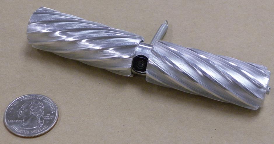
In contrast to the fixed-base miniature robots, mobile robots possess the capability to navigate the abdominal cavity for tissue manipulation and organ exploration. Mobile robots are specifically designed to navigate the smooth and deformable terrain of the abdominal cavity with the help of two independently driven helical-profiled wheels. This wheel design affords sufficient traction without causing any trauma to the tissues. This mobile robot is 15 mm in diameter and 75 mm in length and the safety and mobile capabilities of this robot in the abdominal cavity have been demonstrated in porcine tests[28].
A second generation prototype of this mobile robot, shown in Figure 1, integrated the navigation capability of this robot with an adjustable-focus robotic camera system. The mobile adjustable-focus robotic camera is 20 mm in diameter and has two independently motor-driven wheels and a counter-rotation preventing tail. This system is capable of forward, reverse and turning motion within the abdominal cavity. The capability of this mobile imaging robot to navigate and explore various abdominal organs was tested in an in vivo porcine model[28]. After insertion through a modified laparoscopic port, the mobile camera system safely navigated the abdominal organs and provided a focused view of various abdominal regions. The mobile robotic camera system also provided sole visual feedback and enhanced depth perception during a porcine cholecystectomy. Insertion of such a mobile imaging robot through a standard laparoscopic port could eliminate the need for a separate camera port during abdominal surgeries.
Figure 1 Mobile in vivo imaging robot.
MOBILE IN VIVO BIOPSY ROBOT
A modified prototype of the mobile imaging robot is the mobile in vivo biopsy robot, shown in Figure 2. This in vivo biopsy robot is essentially similar in design to the mobile in vivo imaging robot and possesses independently controlled wheels for mobility, a camera system for surgical imaging and a 2.4 mm wide robotic grasper for biopsy. The biopsy robot is designed to generate sufficient extraction force for tissue biopsy. The mobile imaging camera system on this robot provided visual feedback and helped select a suitable biopsy site. The biopsy graspers were then successfully applied and hepatic tissue biopsy was accomplished[29]. The biopsy robot was then retracted through the entry incision and thus demonstrated successful one-port biopsy and tissue manipulation. This robot provides the added advantage of surgical task assistance and tissue manipulation compared to the previous mobile imaging robots that solely provided visualization assistance during abdominal surgery.
Figure 2 In vivo biopsy robot with biopsy grasper.
MOBILE ENDOLUMINAL ROBOT
The mobile endoluminal robot is 12 mm in diameter and 75 mm long with two independently driven wheels that provide forward, reverse and turning capability in the abdominal cavity. The ability of the mobile endoluminal robot for transgastric exploration under esophagogastroduodenoscopic (EGD) control was successfully demonstrated in porcine models[30,31]. The robot was advanced into the gastric cavity through an overtube placed under EGD control. The robot was introduced into the peritoneal cavity through a transgastric incision performed with an endoscopic needle-knife. A robot-assisted abdominal cavity exploration including liver and small bowel manipulation was successfully completed. Subsequently, the robot was retracted and an endoscopic closure of transgastric incision site was achieved. The robot was eventually retrieved with an endoscopic snare demonstrating the potential ability of these in vivo robots to perform natural orifice surgery.
IN VIVO COOPERATIVE ROBOTS
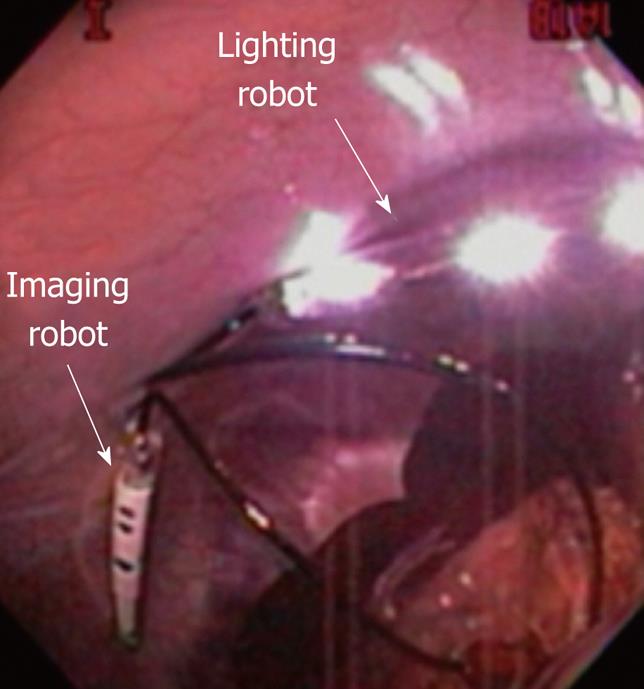
In vivo robots confirm to the geometry of the lumen and their size specifications are in accordance with natural orifice size presenting the potential that a family of miniature in vivo robots could be synchronously deployed in the abdominal cavity for natural orifice surgery. The concept of cooperative robots, shown in Figure 3, each providing spatial orientation and specific task assistance during surgical procedures has been demonstrated in a non-survival porcine model[32]. Three miniature in vivo robots, including a peritoneum-mounted imaging robot, a lighting robot and a retraction robot, are designed for specific surgical tasks. The imaging robot is a 12 mm robot consisting of an outer tube that houses an inner tube with a lens, camera board and three direct current (DC) micromotors for rotation. The robot is fitted with LED for illumination and enables the device to provide video feedback without supplemental light source. The imaging robot can be re-positioned in the abdominal cavity by manipulating external magnetic handles that attract magnets in the robot’s body. This magnetically anchored imaging robot is designed to provide video feedback on a standard monitor during surgical procedures. The lighting robot has an outer tube that houses six white LEDs and is attached to the interior abdominal wall with external magnetic handles. The retraction robot consists of two embedded magnets and a tethered grasping device. A magnetic DC micromotor in the body of the robot coupled with a drum provides rotational movement and activates a grasping device. All three robots are appropriately sized to be inserted through a standard laparoscopic trocar or through a natural orifice during NOTES procedure.
Figure 3 Cooperative robots: Imaging and lighting robot used together.
These three robots in conjunction with a standard upper endoscope have been used in a non-survival porcine NOTES procedure[32]. For this procedure, initially an overtube was placed with the assistance of an endoscope and advanced into the peritoneal cavity. The three robots were then deployed in the peritoneal cavity and magnetically positioned along the upper abdominal wall. In this cooperative procedure, the imaging robot was able to provide high quality video feedback for peritoneal cavity exploration. The retraction robot was specifically used for manipulation of surgical targets such as bowel and gallbladder. Thus, stable imaging by the imaging robot, adequate illumination by the lighting robot and tissue manipulation by the retraction robot provide proof of concept for the use of multiple, independent, specialized robotic devices in a NOTES approach.
IN VIVO DEXTEROUS ROBOT
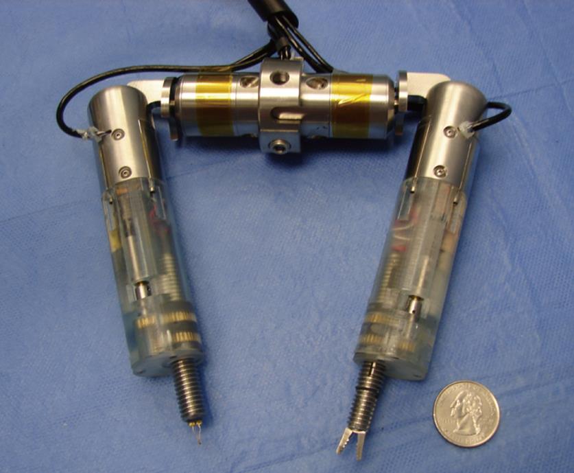
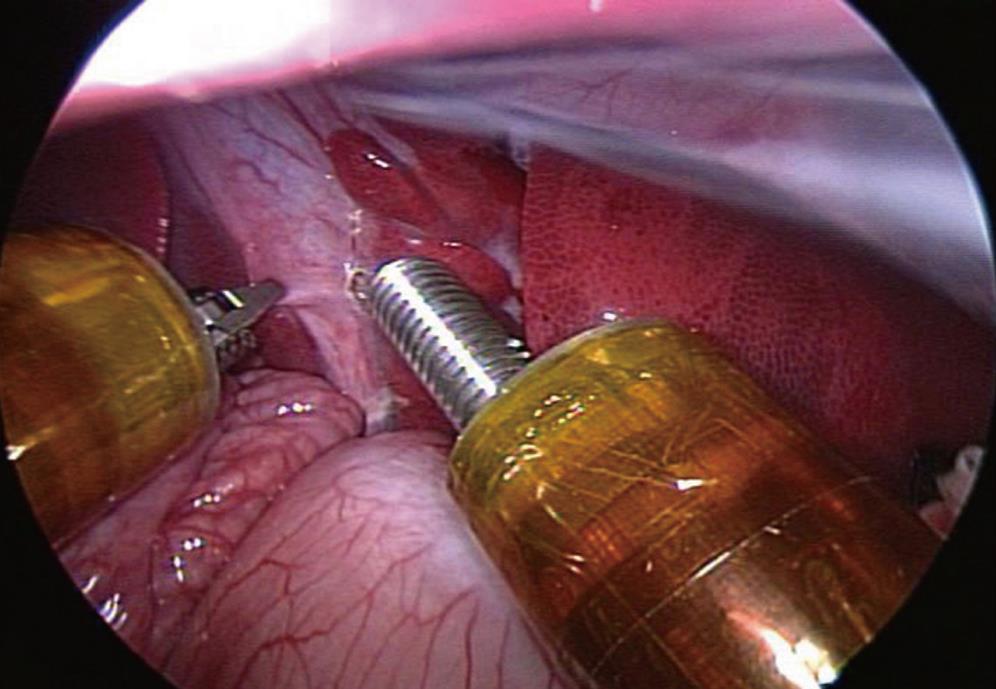
The dexterous miniature in vivo robot for NOTES is a multi-functional robot with ability for tissue retraction and manipulation, stereovision imaging, cautery, and tissue grasping capability. The design of the robot consists of two arms connected to a central body as shown in Figure 4. Each arm consists of upper and lower segments. The upper arm is connected to the central body by a rotational shoulder joint. Retraction and extension is achieved by a lower arm that telescopes in and out of an upper arm. The lower arm is fitted with either a grasper forceps or a cautery end effector. This robot has a remote surgeon interface console. The surgeon control interface is remote in location and comprises of two controllers, a display, and a foot pedal. Each robot arm movement is controlled by the movement of the controllers that are located remotely. Grasper and cautery extensions can be activated when required, at the push of a button. The video feedback from a standard laparoscope is displayed on a screen between the two controllers. Using the surgeon interface console, positioning of the dexterous robot arms with adequate workspace can be performed remotely. Each quadrant of the abdomen can be imaged and surgically accessed through this technique without requiring additional incisions. This robot has been used to perform cholecystectomy and small bowel dissection in a porcine model[33]. This robotic device was endoscopically deployed in the peritoneal cavity through a gastrotomy incision. On-board video feedback from the robot enabled visualization of the small bowel for further manipulation. A small bowel dissection was then performed with the help of forceps on one arm and cautery on the other arm. The small bowel was grasped and retracted with one arm allowing for access of the cautery. The cautery arm was then extended and was able to cauterize the bowel. The positioning of the arms allows for off-axis forces to be applied during tissue retraction and dissection. Once in the peritoneal cavity, this robot provided a stable platform for visualization, dexterous capability to apply off-axis forces, and better triangulation capability for tissue manipulation. Tissue manipulation capability of the in vivo dexterous robot is shown in Figure 5. Presently, the initial prototype of this robot is large and hence an abdominal incision was utilized for adequate access for a cholecystectomy. Further miniaturization of the robot for performing a NOTES procedure in a remote fashion is ongoing.
Figure 4 Prototype in vivo dexterous robot with grasper and cautery end effector arms.
Figure 5 In vivo dexterous robot performing tissue manipulation during porcine cholecystectomy.
NOTES FUTURE PERSPECTIVES
Miniature in vivo robotics is a novel platform and an alternative approach for NOTES. Although successful application of several distinct in vivo robots has been demonstrated in animal models, limitations of their use remain. Currently, some of these robots are best suited for use in conjunction with laparoscopic and endoscopic instruments. To be completely NOTES-compatible, further refinement of these robotic devices is necessary. Nevertheless, successful application of some of these in vivo robots for NOTES has been shown in animal models. For robots that are large in size, further miniaturization is necessary for NOTES approach. Additionally, some of these robots still remain externally tethered for power. Future tether-free applications will be adequately battery-powered and use wireless technology[21]. Development of in vivo robotic assistants with a range of end effectors for specialized complex tasks such as tissue dissection, tissue manipulation and organ retraction, better cauterization, enhanced stereoscopic visualization, surgical suturing capability, and closure of visceral incision endoscopically is necessary for translation into NOTES technology.
Future flexible robotics platform for NOTES will require development of robotic endoscopes with the ability to develop a stable platform for complex surgical tasks without compromising endoscope tip maneuverability. A computer-controlled robotic platform may benefit tasks requiring surgical precision, navigation through peritoneal cavity, and complex maneuvers of the endoscope tip. This computerized robotic platform would allow for surgically precise, accurately controlled and complex maneuvers of the endoscope during surgical procedures. Advances in instrumentation for NOTES would include flexible robotics platform-specific graspers, forceps, scissors, needle-drivers, coagulators and other surgical instruments with easy insertion and quick exchangeability[23,24].
In addition to these robotic platform-specific developments, several issues specific to NOTES such as the optimal site for visceral incision, a standardized technique for NOTES procedures and adequate closure techniques need to be addressed[11]. These issues along with technology development will ultimately determine if the scope of NOTES can be broadened to include complex surgical procedures.
CONCLUSION
A miniature in vivo robotics platform represents a novel and alternative approach for NOTES. Such a platform affords several advantages such as enhanced visualization, better surgical dexterity and significantly improved triangulation capability for NOTES procedures. Development of task-specific in vivo robotic devices for use during NOTES provides enhanced surgical dexterity. Development of a totally intraperitoneal team of robots that cooperatively perform surgical procedures in animal models has been accomplished. Currently, robots that can perform NOTES and other laparoscopic procedures in a remote fashion are being developed. Although these technologies are still in pre-clinical development, a miniature robotics platform provides a unique method for addressing the limitations of minimally invasive surgery, and NOTES in particular.
Peer reviewer: Dr. Simone Ferrero, San Martino Hospital and University of Genoa, Largo R. Benzi 1, Genoa 16131, Italy
S- Editor Li LF L- Editor Hughes D E- Editor Yang C


