Published online Jan 27, 2010. doi: 10.4240/wjgs.v2.i1.32
Revised: August 12, 2009
Accepted: August 19, 2009
Published online: January 27, 2010
Foreign bodies in the rectum wall, whose most common causes are aberrant sexual activity and intake of small fishbone fragments by mistake, usually have a clear history, presenting an acute upset. However, chronic presence of a foreign-body can result in inflammatory reaction and stromal proliferation, and can even accelerate the occurrence and deterioration of tumors through different mechanisms, such as reactive oxygen species. Foreign bodies in the rectum wall may induce complications and lead to misdiagnosis. Colonoscopy and biopsy pathology are one of the most trusted techniques for diagnosis.
- Citation: Kang NS, Qin DP. Cancer-like foreign-body in rectum wall: A case report. World J Gastrointest Surg 2010; 2(1): 32-34
- URL: https://www.wjgnet.com/1948-9366/full/v2/i1/32.htm
- DOI: https://dx.doi.org/10.4240/wjgs.v2.i1.32
A foreign body penetrating into the rectum wall can result in perforation, hemorrhage and infection, etc. Long persistence of a needle-shaped body in the rectum, arriving via ingestion may lead to perianal abscess and archosyrinx. However, intestinal perforation and massive hemorrhage are usually caused by introduction of the foreign body through the anus, and may relate to violence and large volume. Insufficient cognition of the disease may result in misdiagnosis. This case of foreign body in the rectum is really rare because it resulted from defecation over a grassy tussock without the emergency trauma.
A 63-year-old male patient was hospitalized on August 25, 2007. He had suffered tenesmus for two months and flat stool for two weeks. He did not seek any treatments because he had no abdominal pain, abdominal distention, diarrhea, hematochezia or shape or properties changes of stool. In the previous two weeks, he felt defecation had become more difficult, and his stool became pale and accompanied by mucus. He had also lost about 5 kg of weight.
Physical examination revealed a flat and soft abdomen, positive tenderness and rebound tenderness and no mass. Digital rectal examination revealed a hard, fixed mass with hazy borderline, a tender, rough surface about 5 cm beyond the anus edge and bloody dactylotheca. The upper fringe of the mass was untouchable. No supplementary tests had been done.
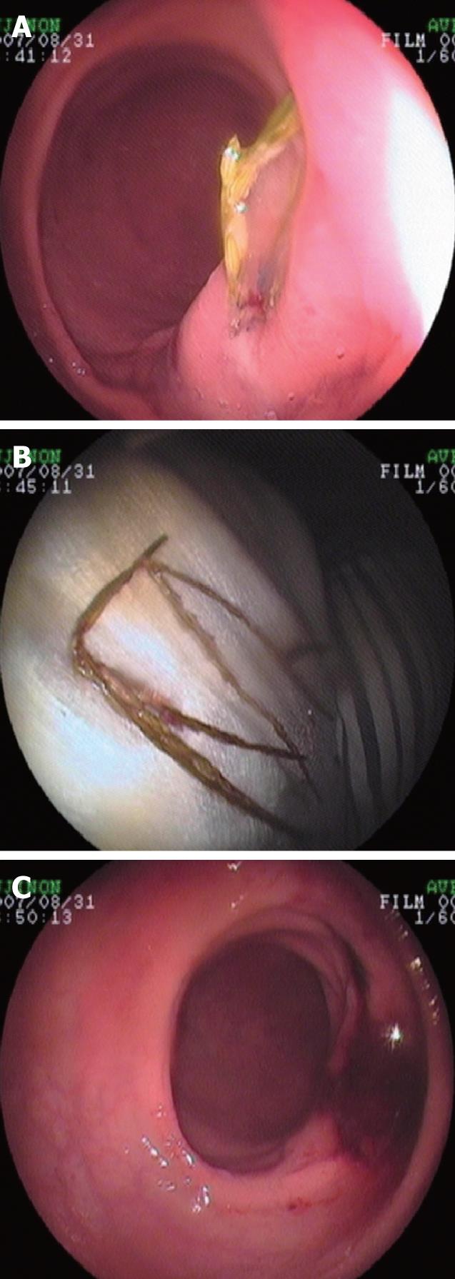
Rectal carcinoma was considered as the original diagnosis. Occult blood was positive. Colonoscopy showed that, 5 cm above anus, there was a ridged and soft tumor with a broken mucous membrane and partial phyllodes change. The endoscopic diagnosis was rectal polyp, and the pathological diagnosis was chronically inflamed mucosa. Tumor markers were all negative. Abdominal CT found local irregular incrassation of the left rear rectum wall accompanied by hazy clearance of ambient fat which was considered to have been caused by rectal carcinoma. Four days later, another colonoscopy was carried out. When the biopsy was being performed, a grass stalk was found in the ridged focus of the rectal front wall about 5 to 6 cm above the anus (Figure 1A). After some grass stalks were removed (Figure 1B), the bulge disappeared (Figure 1C). The endoscopic diagnosis was foreign body of rectal mucosa. Enquiry after the colonoscopy revealed the patient had a history of defecation in over grassy tussocks. Thus the final diagnosis was foreign body in the rectal wall. Follow-up after one year confirmed that the patient was healthy.
Foreign bodies in the rectal wall are rare, and usually relate to foreign bodies in the rectum. The foreign body may come from the mouth or anal canal. There are rare examples of digesting a foreign body, usually caused by intake of small fishbone fragment and other items by mistake which are not arrested or don’t penetrate into all the intestines above the rectum. They finally penetrate into the rectum wall, rectal ampulla or anal canal in particular[1]. This type of insult often has chronic onset or develops into some associated diseases, such as perianal abscess and anal fistula[2]. The majority of bodies entering via the anus route relate to aberrant sexual activity[3,4]. Fragile appliances break and penetrate into the rectum wall during sexual activity. This type of injury usually has acute onset and the original diagnosis can be concluded by enquiry.
Rectum carcinoma is one of the commonly seen tumors of the digestive tract. The incidence rate increases with the age. It is easily ignored in the early stages because of few and unrepresentative symptoms such as occult blood and changes of defecation. The diagnosis depends on associated supplementary tests such as colonoscopy, digital rectal examination and confirmatory pathology.
A polyp-like bulge of the rectal mucosa in this case resulted from stimulation by a foreign body. The mechanism is that the foreign body induces an inflammatory reaction and stromal proliferation, and finally causes foreign-body granuloma which may lead to misdiagnosis[5]. Foreign bodies induce inflammatory reaction, and especially stromal proliferation, where the exogenous materials are incorporated and undigested. Possible factors include inflammatory cytokines, reactive oxygen species, and other particles which act as carriers of carcinogen such as polycyclic aromatic hydrocarbons and so on[6]. Such foreign-body-induced carcinogenesis is also recognized as a step of tumor progression. Tumor development and progression are accelerated inevitably by inflammation resulting from foreign bodies. Reactive oxygen species derived from inflammatory cells are one of the most important genotoxic mediators which accelerate the process[7]. This case is of obvious inducement and acute history, and the symptoms were similar to rectum carcinoma, such as changes in defection, weight loss, positive occult blood, low fever, and the results of digital rectal examination and CT. However, the diagnosis was not supported by the pathology. Therefore, another colonoscopy was carried out. Finally, a foreign body was found and removed, excluding the probability of rectal carcinoma and permitting a correct diagnosis. It will be seen from this investigation that colonoscopy and biopsy pathology play an important role in diagnosing carcinoma of the large intestine and that many factors can causes ridgy change in the rectal mucous membrane. Foreign body in the rectal wall is a possible differential diagnosis.
Peer reviewer: Shuji Nomoto, PhD, Department of Surgery, Graduate School and Faculty of Medicine, Nagoya University, 65 Tsurumai-cho, Showa-ku, Nagoya 466-8550, Japan
S- Editor Li LF L- Editor Lalor PF E- Editor Lin YP
| 1. | Schofield PF. Foreign bodies in the rectum: a review. J R Soc Med. 1980;73:510-513. |
| 2. | Liu XS. Perianal abscess caused by foreign body in rectum: a case report. Dachang Gangmenbing Waike Zazhi. 1997;3:10. |
| 3. | Subbotin VM, Davidov MI, Faĭnshteĭn AV, Abdrashitov RR, Rylov IuL, Sholin NV. [Foreign bodies of the rectum]. Vestn Khir Im I I Grek. 2000;159:91-95. |
| 4. | Cohen JS, Sackier JM. Management of colorectal foreign bodies. J R Coll Surg Edinb. 1996;41:312-315. |
| 5. | Junghans R, Schumann U, Finn H, Riedel U. [Foreign body granuloma of the head of the pancreas caused by a fish bone--a rare differential diagnosis in head of the pancreas tumor]. Chirurg. 1999;70:1489-1491. |
| 6. | Borm PJ, Driscoll K. Particles, inflammation and respiratory tract carcinogenesis. Toxicol Lett. 1996;88:109-113. |
| 7. | Okada F. Beyond foreign-body-induced carcinogenesis: impact of reactive oxygen species derived from inflammatory cells in tumorigenic conversion and tumor progression. Int J Cancer. 2007;121:2364-2372. |


