Published online Feb 27, 2025. doi: 10.4240/wjgs.v17.i2.102354
Revised: December 4, 2024
Accepted: December 16, 2024
Published online: February 27, 2025
Processing time: 98 Days and 22.6 Hours
Acute abdominal pain is one of the most common gastrointestinal symptoms. The etiology of acute abdomen can be challenging for gastroenterologists to establish. Cecal foreign body is a rare cause of cecal perforation.
We report a 35-year-old male from China who initially exhibited symptoms su
We emphasize the importance of a detailed patient history, accurate diagnosis and proper treatment in patients with acute abdomen.
Core Tip: Foreign body ingestion is common in China, but it is rare to cause perforation of the caecum. In cases of complications such as intestinal perforation, particularly when foreign objects are unintentionally ingested, the diagnosis of the etiology of acute abdomen is quite challenging. Doctors should recognize the importance of computed tomography, as it is crucial for their diagnosis and subsequent treatment. After an object has been ingested, the recommended first-line treatment is to remove it endoscopically. Patients should receive appropriate intervention to prevent complications.
- Citation: Chen T. Acute abdominal pain complicated by cecal perforation caused by an unnoticed swallowed toothpick: A case report. World J Gastrointest Surg 2025; 17(2): 102354
- URL: https://www.wjgnet.com/1948-9366/full/v17/i2/102354.htm
- DOI: https://dx.doi.org/10.4240/wjgs.v17.i2.102354
Acute abdominal pain is the most common reason for admission to the department of emergency gastroenterology[1]. The most common cause of acute abdomen is intestinal disease, and the incidence of acute abdomen is 4%-5%[2]. The main causes of acute abdomen are acute appendicitis, followed by acute intestinal obstruction, acute cholecystitis and hollow organ perforation[1]. Perforation of the cecum due to toothpick ingestion is rare, and its clinical features are similar to appendicitis, which can present a diagnostic challenge. There are only few cases published in the literature[3]. In this report, we present the case of a 35-year-old male from China who initially presented with symptoms suggestive of acute appendicitis. Combined with the patient's recent history of eating fish and computed tomography (CT) results, the cecal perforation caused by a fishbone was preliminarily considered. However, during colonoscopy, a toothpick was identified as the underlying cause of the patient's potential presentation. We clarified the diagnosis of foreign body through appropriate diagnostic methods, and successfully removed the toothpick through minimally invasive treatment to avoid misdiagnosis of laparotomy.
A 35-year-old man was admitted to our hospital with a 2 days history of right lower abdominal pain and low-grade fever.
The pain was recently aggravating, dull-aching, intermittent, lasting for hours, and not related to food intake.
The patient was healthy in the past.
The patient's history of smoking, alcohol consumption and surgery, as well as, family history, development history and social history and systematic examinations were unremarkable.
His vital signs were normal, except for a central body temperature of 37.8 °C. Physical examination suggested mild pain with positive rebound tenderness in the right lower abdominal quadrant.
Blood counts were normal, but plasma high-sensitivity C-reactive protein was increased at 34.06 mg/dL.
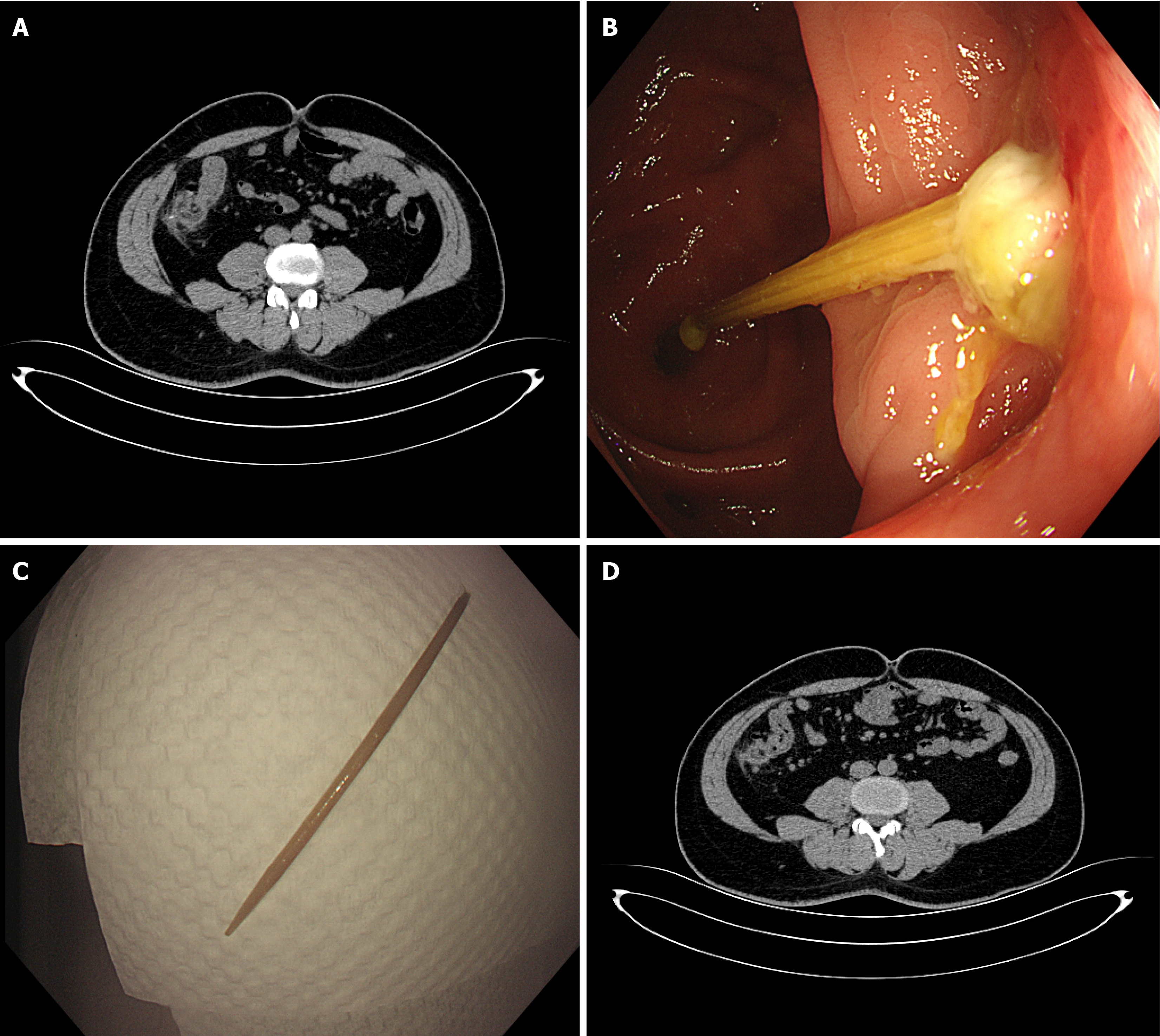
Abdominal CT revealed a strip of high-density shadows protruding beyond the intestinal cavity outline, with a small amount of peritoneal seepage in the ileocecal area (Figure 1A). After further questioning of the patient, we learned that the patient had a history of eating fish 2 days before. Combined with medical history, the possibility of perforation of foreign body in fishbone and peripheral peritonitis was considered. After digestive endoscopy and surgical consultation, endoscopic removal of the objects was suggested. The patient underwent colonoscopy, which to our surprise, revealed a wooden toothpick of 6.5 cm in length stuck in the ileocecum (Figure 1B). The patient was oblivious to the fact that he had deliberately swallowed the toothpick, but he did recall using it while intoxicated during a dinner with friends a few nights ago.
Cecal perforation caused by an unnoticed swallowed toothpick.
The wooden toothpick was removed via a minimally invasive procedure using a foreign body forceps under colonoscopy (Figure 1C).
Before discharge, the patient was examined by abdominal CT that showed a small amount of peritoneal exudation in the ileocecal area, which was significantly reduced compared with before, and the original foreign body was not seen (Figure 1D). The patient did not complain of abdominal pain, and there was no abdominal tenderness. The patient made an uneventful recovery and was discharged within 5 days following treatment.
We report on a young man who presented primarily with acute abdominal pain in the lower right region and whose blood tests indicated elevated inflammatory markers. Danish et al[1] reported that acute appendicitis is the leading cause of acute abdomen, accounting for half of total admissions. Therefore, we initially thought that acute abdominal pain was more likely to be associated with acute appendicitis. CT diagnosis of acute appendicitis is highly accurate, with a reported sensitivity between 90% and 100%[2]. Hence, it is indispensable for the patient to undertake abdominal CT for further confirmation of the diagnosis. However, an elongated foreign body in the ileocecal area was identified on the CT scan, as were signs of intestinal perforation, including an image of the foreign body passing through the intestinal wall, and increased mesenteric fat density.
Although foreign body ingestion is common, it mostly occurs in children[4]. Even in adults, foreign body ingestions are usually due to mental illness, poisoning, or secondary gain, which are usually intentional[5]. It has been reported that most cases of accidental ingestion of foreign bodies are caused by dentures[6]. Toothpick ingestion is a rare event that results in serious gut injuries with perforation[3]. As a result, patients often do not report to their doctors that they have ingested a foreign body, potentially complicating and delaying diagnosis. Even though we confirmed that the patient's abdominal pain was attributed to a foreign body and hypothesized that the cecal perforation was induced by a fish bone, we ultimately discovered that the foreign body was actually a toothpick, although the treatment remained the same.
Previous literature has reported that male sex, toothpick chewing habits, toothpick-containing diet and alcohol consumption are the main risk factors associated with toothpick intake[3]. Most toothpicks are swallowed unintentionally[3,7]. Sharp and elongated foreign bodies are more likely to lead to complications such as gastrointestinal perforation. Perforation can occur anywhere in the gastrointestinal tract, but is most common in areas that are physiologically an
CT is the best imaging method to identify foreign bodies, and it can determine the exact location of the perforation site and provide a basis for subsequent treatment[5]. Emergency laparotomy is necessary when the foreign body is sharp or cannot be removed endoscopically, or when the patient presents with acute abdomen and intestinal perforation[8]. Due to the fact that intestinal perforation is brought about by impaction and the progressive erosion of foreign bodies in contact with the intestinal wall, the perforation site is typically covered by fibrin, omentum and other intestinal loops, thereby restricting the entry of a large amount of gas into the abdominal cavity[9]. Foreign bodies ought to be eliminated whe
This was a single case, without long-term follow-up, which makes it difficult to generalize results.
Foreign body ingestion is common in China, but it is rare to cause perforation of the cecum. In cases of complications such as intestinal perforation, particularly when foreign objects are unintentionally ingested, it is challenging to establish the etiology. Doctors should recognize the importance of CT, as it is crucial for diagnosis and subsequent treatment. After an object has been ingested, the recommended first-line treatment is to remove it endoscopically. Patients should receive appropriate intervention to prevent complications. At the same time, patient education is also important in the pre
| 1. | Danish A. A retrospective case series study for acute abdomen in general surgery ward of Aliabad Teaching Hospital. Ann Med Surg (Lond). 2022;73:103199. [RCA] [PubMed] [DOI] [Full Text] [Full Text (PDF)] [Cited by in Crossref: 2] [Cited by in RCA: 1] [Article Influence: 0.3] [Reference Citation Analysis (0)] |
| 2. | Priola AM, Priola SM, Volpicelli G, Giraudo MT, Martino V, Fava C, Veltri A. Accuracy of 64-row multidetector CT in the diagnosis of surgically treated acute abdomen. Clin Imaging. 2013;37:902-907. [RCA] [PubMed] [DOI] [Full Text] [Cited by in Crossref: 6] [Cited by in RCA: 11] [Article Influence: 0.9] [Reference Citation Analysis (0)] |
| 3. | Lovece A, Asti E, Sironi A, Bonavina L. Toothpick ingestion complicated by cecal perforation: case report and literature review. World J Emerg Surg. 2014;9:63. [RCA] [PubMed] [DOI] [Full Text] [Full Text (PDF)] [Cited by in Crossref: 9] [Cited by in RCA: 14] [Article Influence: 1.3] [Reference Citation Analysis (0)] |
| 4. | Gumbs S, Ausqui G, Orach T, Ramcharan A, Donaldson B. An Unusual Case of Cecal Perforation: Accidental Ingestion of a Tooth in an Elderly Trauma Patient. Cureus. 2023;15:e45467. [RCA] [PubMed] [DOI] [Full Text] [Reference Citation Analysis (0)] |
| 5. | Nicolodi GC, Trippia CR, Caboclo MF, de Castro FG, Miller WP, de Lima RR, Tazima L, Geraldo J. Intestinal perforation by an ingested foreign body. Radiol Bras. 2016;49:295-299. [RCA] [PubMed] [DOI] [Full Text] [Full Text (PDF)] [Cited by in Crossref: 24] [Cited by in RCA: 37] [Article Influence: 4.1] [Reference Citation Analysis (0)] |
| 6. | Goh BK, Tan YM, Lin SE, Chow PK, Cheah FK, Ooi LL, Wong WK. CT in the preoperative diagnosis of fish bone perforation of the gastrointestinal tract. AJR Am J Roentgenol. 2006;187:710-714. [RCA] [PubMed] [DOI] [Full Text] [Cited by in Crossref: 162] [Cited by in RCA: 136] [Article Influence: 7.2] [Reference Citation Analysis (0)] |
| 7. | Yao Y, Yan G, Feng L. A Patient with Acute Abdominal Pain Caused by an Unnoticed Swallowed Toothpick Misdiagnosed as Acute Appendicitis. Arch Iran Med. 2022;25:274-276. [RCA] [PubMed] [DOI] [Full Text] [Full Text (PDF)] [Cited by in Crossref: 4] [Cited by in RCA: 4] [Article Influence: 1.3] [Reference Citation Analysis (0)] |
| 8. | Zarei M, Shariati B, Bidaki R. Intestinal Perforation Due to Foreign Body Ingestion in a Schizophrenic Patient. Int J High Risk Behav Addict. 2016;5:e30127. [RCA] [PubMed] [DOI] [Full Text] [Full Text (PDF)] [Cited by in Crossref: 5] [Cited by in RCA: 5] [Article Influence: 0.6] [Reference Citation Analysis (0)] |
| 9. | Pinero Madrona A, Fernández Hernández JA, Carrasco Prats M, Riquelme Riquelme J, Parrila Paricio P. Intestinal perforation by foreign bodies. Eur J Surg. 2000;166:307-309. [RCA] [PubMed] [DOI] [Full Text] [Cited by in Crossref: 109] [Cited by in RCA: 133] [Article Influence: 5.3] [Reference Citation Analysis (0)] |
| 10. | Ibrahim AF, Hussen MS, Tekle Y, Mohammed H. A rare case of cecal foreign body leading to cecal perforation in 12-year-old child: a clinical case report and review of literature. Ann Med Surg (Lond). 2024;86:1676-1680. [RCA] [PubMed] [DOI] [Full Text] [Full Text (PDF)] [Reference Citation Analysis (0)] |


