Published online Feb 27, 2024. doi: 10.4240/wjgs.v16.i2.518
Peer-review started: December 12, 2023
First decision: January 2, 2024
Revised: January 13, 2024
Accepted: January 24, 2024
Article in press: January 24, 2024
Published online: February 27, 2024
Processing time: 75 Days and 6.7 Hours
Gastric cancer is a leading cause of cancer-related deaths worldwide. Prognostic assessments are typically based on the tumor-node-metastasis (TNM) staging system, which does not account for the molecular heterogeneity of this disease. LATS2, a tumor suppressor gene involved in the Hippo signaling pathway, has been identified as a potential prognostic biomarker in gastric cancer.
To construct and validate a nomogram model that includes LATS2 expression to predict the survival prognosis of advanced gastric cancer patients following ra
A retrospective analysis of 245 advanced gastric cancer patients from the Fourth Hospital of Hebei Medical University was conducted. The patients were divided into a training group (171 patients) and a validation group (74 patients) to deve
The model demonstrated a high predictive accuracy with C-indices of 0.829 in the training set and 0.862 in the validation set. Area under the curve values for three-year and five-year survival prediction were significantly robust, suggesting an excellent discrimination ability. Calibration plots confirmed the high concordance between the predictions and actual survival outcomes.
We developed a nomogram model incorporating LATS2 expression, which significantly outperformed conventional TNM staging in predicting the prognosis of advanced gastric cancer patients postsurgery. This model may serve as a valuable tool for individualized patient management, allowing for more accurate stratification and im
Core Tip: This study focuses on developing a prognostic model for patients with advanced gastric cancer postsurgery by integrating LATS2 expression and clinicopathological features into a nomogram. It highlights the significance of the LATS2 gene in improving prognostic predictions beyond traditional tumor-node-metastasis staging. Our model demonstrates excellent predictive accuracy and clinical utility, which indicates its potential for enhancing individualized patient care through better risk stratification. Future studies should focus on external validation to confirm the model’s applicability across diverse patient populations.
- Citation: Sun N, Tan BB, Li Y. Nomogram model including LATS2 expression was constructed to predict the prognosis of advanced gastric cancer after surgery. World J Gastrointest Surg 2024; 16(2): 518-528
- URL: https://www.wjgnet.com/1948-9366/full/v16/i2/518.htm
- DOI: https://dx.doi.org/10.4240/wjgs.v16.i2.518
Gastric cancer ranks fifth and fourth among all malignant tumors globally in terms of incidence and mortality, res
Nomograms have been used to predict the survival prognosis of cancer patients through the construction of mul
Large tumor suppressor kinase (LATS) was first discovered in Drosophila melanogaster in 1995. An ortholog was found to exist in humans in subsequent studies, which included LATS1 and LATS2[12]. LATS is a core member of the Hippo signaling pathway and plays an important role in maintaining cellular homeostasis[13,14]. LATS also has a role in the development of gastric cancer by inhibiting tumor cell proliferation and promoting apoptosis[15,16].
In this study, 245 gastric cancer patients were retrospectively analyzed to determine the clinical value of LATS2 ex
A total of 245 patients with advanced gastric cancer who underwent surgery in the Department of Surgery at the Fourth Affiliated Hospital of Hebei Medical University from March 2015 to March 2017 were retrospectively selected. The patients were randomly divided into a training group and a validation group at a 7:3 ratio. The training group was used to screen variables and construct models, whereas the validation group was used to validate the models obtained from the training group. The tumor were surgically resected and analyzed for pathology and by immunohistochemical staining. The tumor-node-metastasis (TNM) stage was determined according to the AJCC 8th edition standard TNM staging system. The data in were obtained from the postoperative pathology reports and the patient clinical information was collected through the hospital’s independent information system. For immunohistochemistry, the intensity and extent of staining were assessed semi-quantitatively, with scores of 0-3 defined as low expression and scores of 4-9 defined as high expression. This was a retrospective study approved by the Ethics Committee of the Fourth Hospital of Hebei Medical University (Approval No. 2019ME0039).
Inclusion criteria: (1) Confirmed diagnosis of advanced gastric cancer along with radical gastrectomy (D2 lymph node dissection, R0 resection); (2) M0 patients without distant metastasis; (3) Diagnosis of primary gastric adenocarcinoma, which was clearly defined by postoperative pathology and immunohistochemical staining; (4) Complete medical records and immunohistochemistry analysis data; (5) No chemotherapy, radiotherapy, immunotherapy, or other tumor present; and (6) The depth of cancer cell invasion reached the muscle layer, serous layer, or extraserous layer (T stage ≥ T2).
Exclusion criteria: (1) Patients aged < 18 years old and survival time < 1 month; (2) Combination of severe anemia, infection, autoimmune disease, hematologic disease, or inflammation in the last month; (3) Combination of severe brain, heart, liver, kidney, or other vital organ disease; (4) Recurrence, combination of distant metastasis, or history of other malignant neoplasms; (5) Recent use of antibiotics, anti-inflammatory drugs; and (6) Incomplete pathological data or postoperative loss of patient contact.
The collected clinical data included: gender, age, comorbidities (hypertension, diabetes mellitus, chronic lung disease), history of smoking, LATS2, history of alcohol consumption, tumor size, lymphatic invasion, T-stage, and N stage.
Follow-up data were obtained from inpatient medical records, outpatient medical records, and telephone consultations. Patients were monitored every three months during the first two years following surgery and every six months after two years. Overall survival (OS) was defined as the time from the start of treatment to death from any cause. For patients lacking study endpoints, we recorded the latest follow-up date, which was January 31, 2022, for the included patients. Local recurrence or distant metastasis was determined based on a computed tomography (CT)-enhanced scan, fiberoptic gastroscopy biopsy, bone scan, magnetic resonance imaging, or positron emission tomography/CT.
Categorical variables were expressed as numbers and percentages, and continuous variables were expressed as the median and interquartile range. Differences in the distribution of categorical variables between the training and va
A total of 245 patients were randomly divided into a training group (171) and a validation group (74) in a 7:3 ratio. Among all patients, the majority were male (80.41%) and most of them were older than 65 years (60.00%) (Table 1). More than half of the patients did not have hypertension (64.90%), diabetes mellitus (90.61%), or chronic lung disease (96.33%), whereas a minority had no history of smoking (8.98%). Most of the patients exhibited lymphatic invasion (81.63%) and high LATS2 expression (66.90%). Approximately half of the patients had a history of alcohol consumption. Most patients had a tumor diameter greater than 5 cm (83.67%) and staged as T4 (72.24%) and N3 (30.61%).
| Characteristics | All patients cases (%)/median (IQR) | Training group cases (%)/median (IQR) | Validation group cases (%)/median (IQR) | P value |
| Sex | ||||
| Male | 197 (80.41) | 142 (83.04) | 55 (74.32) | 0.16 |
| Female | 48 (19.59) | 29 (16.96) | 19 (25.68) | |
| Age | ||||
| < 65 | 98 (40.00) | 64 (37.43) | 34 (45.95) | 0.27 |
| ≥ 65 | 147 (60.00) | 107 (62.57) | 40 (54.05) | |
| Hypertension | ||||
| No | 159 (64.90) | 115 (67.25) | 44 (59.46) | 0.30 |
| Yes | 86 (35.10) | 56 (32.75) | 30 (40.54) | |
| Diabetes | ||||
| Yes | 23 (9.39) | 13 (7.60) | 10 (13.51) | 0.22 |
| No | 222 (90.61) | 158 (92.40) | 64 (86.49) | |
| Chronic lung disease | ||||
| Yes | 9 (3.67) | 6 (3.51) | 3 (4.05) | 1 |
| No | 236 (96.33) | 165 (96.49) | 71 (95.95) | |
| Smoking | ||||
| Yes | 223 (91.02) | 154 (90.06) | 69 (93.24) | 0.58 |
| No | 22 (8.98) | 17 (9.94) | 5 (6.76) | |
| Lymphatic invasion | ||||
| Yes | 200 (81.63) | 140 (81.87) | 60 (81.08) | 1 |
| No | 45 (18.37) | 31 (18.13) | 14 (18.92) | |
| LATS2 | ||||
| High | 164 (66.90) | 121 (70.70) | 43 (58.10) | 0.60 |
| Low | 81 (33.10) | 50 (29.30) | 31 (41.80) | |
| Alcohol consumption | ||||
| Yes | 135 (55.10) | 93 (54.39) | 42 (56.76) | 0.84 |
| No | 110 (44.90) | 78 (45.61) | 32 (43.24) | |
| Tumor size | ||||
| > 5 cm | 205 (83.67) | 145 (84.80) | 60 (81.08) | 0.59 |
| ≤ 5 cm | 40 (16.33) | 26 (15.20) | 14 (18.92) | |
| T stage | ||||
| 2 | 45 (18.37) | 26 (15.20) | 19 (25.68) | 0.11 |
| 3 | 23 (9.39) | 15 (8.77) | 8 (10.81) | |
| 4 | 177 (72.24) | 130 (76.03) | 47 (63.51) | |
| N stage | ||||
| 0 | 74 (30.20) | 54 (31.58) | 20 (27.03) | 0.62 |
| 1 | 45 (18.37) | 28 (16.37) | 17 (22.97) | |
| 2 | 51 (20.82) | 35 (20.47) | 16 (21.62) | |
| 3 | 75 (30.61) | 54 (31.58) | 21 (28.38) |
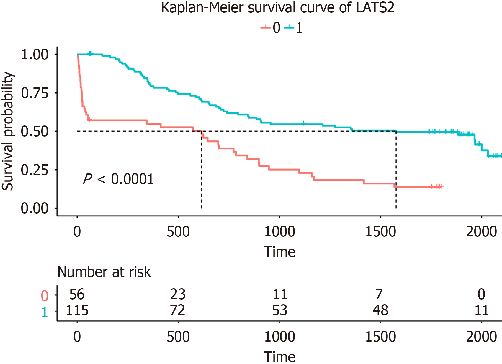
Based on a unifactorial Cox analysis, age, lymphatic invasion, LATS2 expression, tumor size, T stage, and N stage had predictive value (Table 2). In a multifactorial Cox analysis, all of the above factors had independent predictive value, except for age, which was included in the nomogram model. As shown in Figure 1, the survival rate of patients with high LATS2 expression was higher compared with that of low-expressing patients.
| Characteristics | Univariate analysis | Multivariate analysis | |||||
| HR | 95%CI | P value | HR | 95%CI | P value | ||
| Sex | |||||||
| Male | Reference | ||||||
| Female | 0.700 | 0.397-1.233 | 0.217 | ||||
| Age | |||||||
| < 65 | Reference | Reference | 0.199 | ||||
| ≥ 65 | 6.632 | 4.026-10.926 | < 0.001 | 1.780 | 0.739-4.286 | ||
| Hypertension | |||||||
| No | 1.202 | 0.800-1.812 | 0.378 | ||||
| Yes | Reference | ||||||
| Diabetes | |||||||
| Yes | 1.256 | 0.608-2.594 | 0.537 | ||||
| No | Reference | ||||||
| Chronic lung disease | |||||||
| Yes | 0.807 | 0.255-2.552 | 0.716 | ||||
| No | Reference | ||||||
| Smoking | |||||||
| Yes | 0.900 | 0.604-1.312 | 0.605 | ||||
| No | Reference | ||||||
| Lymphatic invasion | |||||||
| Yes | 13.121 | 7.840-21.959 | < 0.001 | 3.251 | 1.519-6.960 | 0.002 | |
| No | Reference | Reference | |||||
| LATS2 | |||||||
| High | 0.327 | 0.217-0.495 | < 0.001 | 0.522 | 0.307-0.887 | 0.016 | |
| Low | Reference | Reference | |||||
| Alcohol consumption | |||||||
| Yes | 1.186 | 0.796-1.767 | 0.402 | ||||
| No | Reference | ||||||
| Tumor size | |||||||
| > 5 cm | Reference | Reference | |||||
| ≤ 5 cm | 319.955 | 42.319-2419.010 | < 0.001 | 79.564 | 8.657-731.251 | < 0.001 | |
| T stage | |||||||
| 2 | Reference | ||||||
| 3 | 6.691 | 2.407-18.598 | < 0.001 | 9.620 | 3.390-27.300 | < 0.001 | |
| 4 | 5.452 | 2.372-12.536 | < 0.001 | 4.156 | 1.753-9.850 | < 0.001 | |
| N stage | |||||||
| 0 | Reference | ||||||
| 1 | 1.650 | 0.824-3.304 | 0.158 | 1.762 | 0.872-3.563 | 0.115 | |
| 2 | 2.237 | 1.186-4.220 | 0.013 | 2.248 | 1.155-4.376 | 0.017 | |
| 3 | 4.332 | 2.452-7.653 | < 0.001 | 3.874 | 2.127-7.056 | < 0.001 | |
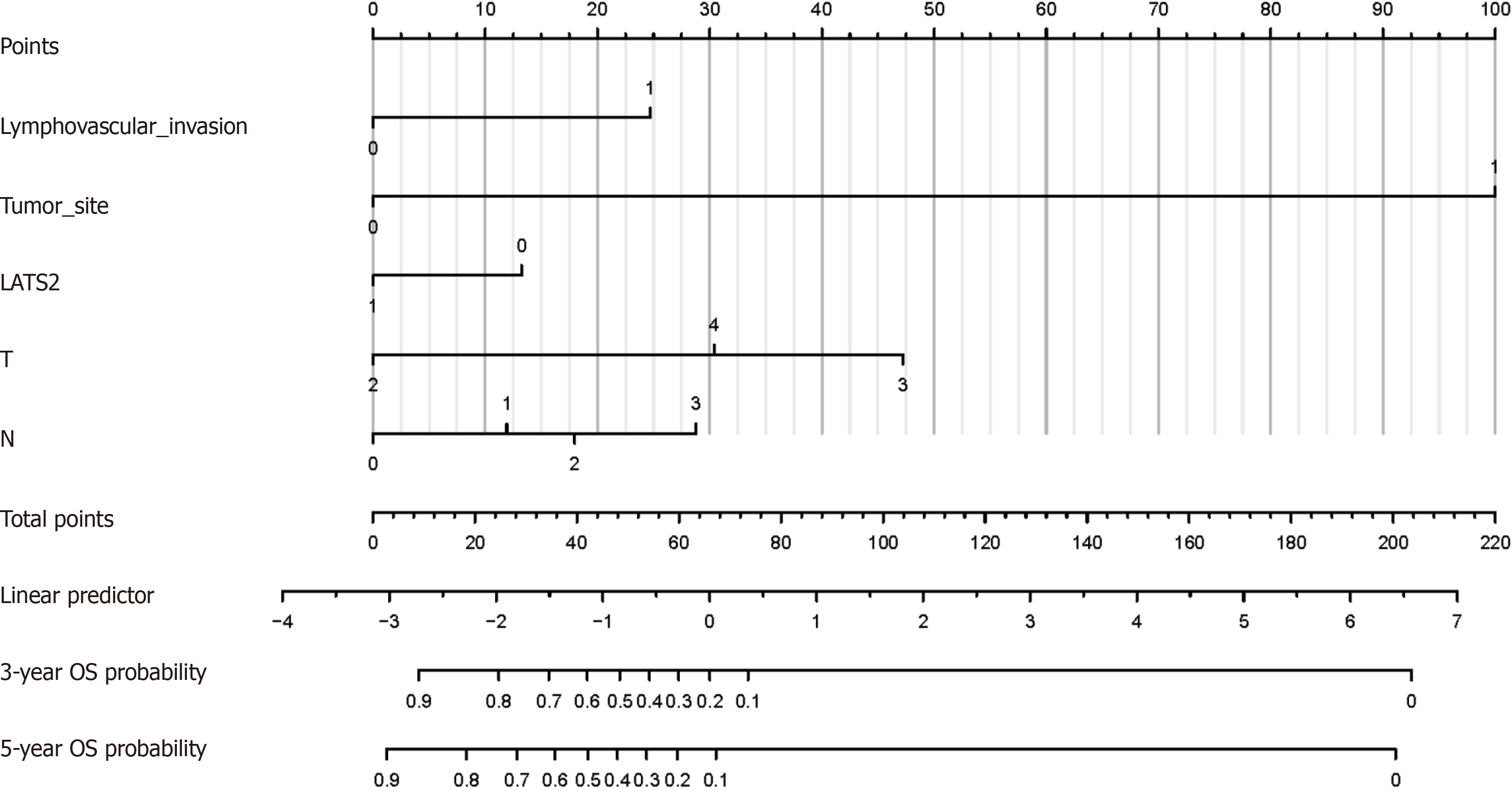
Based on the results of a multifactorial Cox analysis, lymphatic invasion, LATS2, tumor size, T, and N were included in the nomogram (Figure 2), and the consistency indices (C-index) of the three clinical prediction models, namely TN staging, TN staging combined with LATS2, and the nomogram model, were calculated, respectively, according to the results of the clinical data. After comparing the three clinical prediction models, we concluded that the nomogram model had accurate clinical prognostic prediction (Table 3). The C-indexes of the training and validation sets were 0.829 and 0.862, respectively, which were higher compared with those of the two models, TN staging, and TN staging combined with LATS2. The three-year and five-year AUC of TN staging combined with LATS2 in the training set were higher compared with those of the TN staging model, indicating that LATS2 has a positive significance for the prognosis of progressive gastric cancer. The three-year and five-year AUC of the training set of the nomogram graph model were 0.8723 and 0.8525, respectively, which were higher compared with those of the two models of TN staging and TN staging combined with LATS2, which suggests that our model has a good predictive performance (Figure 3). Similarly, the three-year and five-year AUC of the validation set nomogram model were 0.9129 and 0.8763, respectively (Figure 4), indicating that it had a good discriminatory ability. As shown in Figures 5 and 6, the prognosis predicted by the nomogram model was in high agreement with the actual real-world situation.
| Variables | Training group | Validation group | |||
| C-index | 95%CI | C-index | 95%CI | ||
| Overall survival | TN stage | 0.702 | 0.653-0.751 | 0.756 | 0.682-0.830 |
| TN stage + LATS2 | 0.785 | 0.742-0.828 | 0.830 | 0.761-0.899 | |
| Nomogram | 0.829 | 0.788-0.870 | 0.862 | 0.805-0.919 | |
Accurate prediction of postoperative prognosis is important for optimizing treatment strategies and improving outcomes in patients with advanced gastric cancer. In this study, we developed and validated a prognostic model incorporating LATS2 expression and clinicopathological characteristics to predict overall survival at three and five years following radical surgery for advanced gastric cancer.
Several findings indicate that our nomogram model has significant clinical value for postoperative prognosis prediction. First, the model showed good discrimination, with C-indices of 0.829 and 0.862 in the training and validation sets, respectively. The AUCs at three and five years were also high, ranging from 0.8525 to 0.9129, further demonstrating the strong predictive accuracy. Calibration plots revealed a high level of agreement between the predicted and actual survival. In addition, a decision curve analysis showed that the predictions using our model resulted in a superior net benefit compared with predictions based on TNM staging alone or TNM combined with LATS2 across a wide range of threshold probabilities. Taken together, the results provide solid evidence that the model can accurately stratify patients by prognostic risk.
Of note, the incorporation of LATS2 expression data improved model performance over TNM staging alone. High LATS2 expression was associated with a significantly better prognosis, which is consistent with its known tumor suppressive function. As an important component of the Hippo signaling pathway, which regulates cell proliferation and apoptosis, dysregulation of LATS2 affects key oncogenic mechanisms in gastric cancer. Our findings indicate that LATS2 merits inclusion as a prognostic biomarker, lending additional risk stratification information beyond standard clini
In clinical practice, TNM staging is primarily used to roughly predict prognosis; however, it lacks individualization and accuracy in predicting postoperative treatment and prognosis[17]. The construction of effective prognostic tools is of great significance for the individualized treatment of patients, for whom the nomogram has attracted much attention in recent years because of its convenient and intuitive clinical application[18]. Some models for predicting patient prognosis in gastric cancer have been reported, such as a gastric cancer prognosis model based on oxidative stress genes[19], and a model related to scorched death[20]; however. there are many candidate genes involved and research is immature, so the clinical feasibility is not high. For example, age, race, marital status, TNM stage, surgery, chemotherapy, grade, and the number of positive regional nodes can be used to predict the survival of gastric cancer patients[21]; however, the C-index and area under the ROC curve are low, thus the accuracy of prognosis for gastric cancer is not high. To more accurately predict the prognosis of patients with gastric cancer following surgery and for clinical application, it is significant to combine biomarkers that have an important role in gastric cancer with clinicopathological features.
The user-friendly visual format of the nomogram facilitates its application to clinical practice. By mapping the profiles of individual patients onto the diagram, physicians can readily estimate three- and five-year survival probabilities. This may facilitate important management decisions, such as selecting high-risk patients likely to benefit from adjuvant chemotherapy or more intensive follow-up. Dynamic risk assessment at successive time points may also help to de
By enabling more accurate risk stratification and prognosis prediction, this model has the potential to significantly improve clinical decision-making and outcomes for patients undergoing radical surgery for advanced gastric cancer. The next step will be an external validation of the model using patient cohorts from other centers. Broader validation will provide further support for its generalizability and clinical utility. The LATS gene family is a core component of the Hippo pathway and an important regulator of homeostasis in vivo, of which LATS2 expression is low in gastric cancer tissues[16]. Previous studies have confirmed that in gastric cancer, LATS2 is involved in tumor cell growth, invasion, migration[22-24], and mesenchymal transformation[25]. Previous studies have confirmed that LATS2 expression in gas
Some limitations should be acknowledged when interpreting our results. First, the sample size, though adequately powered for model development and internal validation, was relatively small and derived from a single center. Thus, large prospective studies are warranted to validate these findings. Second, data on certain potential prognostic variables, such as the Lauren classification and Borrmann classification, were not available for inclusion. Future models in
In conclusion, we developed an internally validated, robust prognostic model for advanced gastric cancer after radical surgery, incorporating both tumor biomarkers and clinicopathological data. The results indicate that the model enables superior discrimination of low- vs high-risk patients compared with standard prognostic approaches. Following additional external validation, translation of this prognostic tool into clinical practice may significantly assist therapeutic decision-making and ultimately improve patient outcomes. The next steps will be to expand validation of the model across multiple centers as well as investigator-initiated trials to evaluate its impact on clinical management and survival.
Gastric cancer is a significant global health concern, ranking fifth in incidence and fourth in mortality among all cancers. The prognosis is often poor due to late-stage diagnosis. Molecular signaling pathways and gene mutations, like those involving the LATS gene, play a crucial role in the pathogenesis of gastric cancer, affecting prognosis and treatment options.
There is a need for more accurate prognostic models for advanced gastric cancer that can incorporate molecular bio
The objective of this research is to construct a nomogram model based on LATS2 expression and evaluate its predictive accuracy for the survival prognosis of patients with advanced gastric cancer post-surgery.
The study retrospectively analyzed clinical data of 245 advanced gastric cancer patients, dividing them into a training group and a validation group. Univariate and multivariate Cox regression analyses were used to assess the prognostic value of LATS2 expression. The model's performance was analyzed through various statistical methods including C-index, receiver operating characteristic curves, calibration curves, and decision curves.
The nomogram model demonstrated high C-indexes and area under curve values, indicating strong predictive accuracy. Calibration plots showed high agreement between predicted and actual survival, and decision curves indicated the model's superior net benefit over tumor-node-metastasis (TNM) staging alone.
The nomogram model incorporating LATS2 expression provided significant clinical value in predicting the postoperative prognosis of advanced gastric cancer patients. It showed superior discrimination and net clinical benefit compared to TNM staging alone.
The study suggests that the developed model can assist in clinical decision-making, but acknowledges limitations such as the small, single-center sample size. Future research should aim at external validation and include more comprehensive clinical and molecular data to optimize prognostic accuracy.
Provenance and peer review: Unsolicited article; Externally peer reviewed.
Peer-review model: Single blind
Specialty type: Oncology
Country/Territory of origin: China
Peer-review report’s scientific quality classification
Grade A (Excellent): 0
Grade B (Very good): 0
Grade C (Good): C
Grade D (Fair): 0
Grade E (Poor): 0
P-Reviewer: Herrera-Pariente C, Spain S-Editor: Gong ZM L-Editor: A P-Editor: Xu ZH
| 1. | Hirata Y, Noorani A, Song S, Wang L, Ajani JA. Early stage gastric adenocarcinoma: clinical and molecular landscapes. Nat Rev Clin Oncol. 2023;20:453-469. [RCA] [PubMed] [DOI] [Full Text] [Cited by in Crossref: 17] [Cited by in RCA: 32] [Article Influence: 16.0] [Reference Citation Analysis (0)] |
| 2. | Guan WL, He Y, Xu RH. Gastric cancer treatment: recent progress and future perspectives. J Hematol Oncol. 2023;16:57. [RCA] [PubMed] [DOI] [Full Text] [Cited by in RCA: 411] [Reference Citation Analysis (5)] |
| 3. | Gilhaus K, Cepok C, Kamm D, Surmann B, Nedvetsky PI, Emich J, Sundukova A, Saatkamp K, Nüsse H, Klingauf J, Wennmann DO, George B, Krahn MP, Pavenstädt HJ, Vollenbröker BA. Activation of Hippo Pathway Damages Slit Diaphragm by Deprivation of Ajuba Proteins. J Am Soc Nephrol. 2023;34:1039-1055. [RCA] [PubMed] [DOI] [Full Text] [Cited by in Crossref: 4] [Cited by in RCA: 12] [Article Influence: 6.0] [Reference Citation Analysis (0)] |
| 4. | Wang CH, Baskaran R, Ng SS, Wang TF, Li CC, Ho TJ, Hsieh DJ, Kuo CH, Chen MC, Huang CY. Platycodin D confers oxaliplatin Resistance in Colorectal Cancer by activating the LATS2/YAP1 axis of the hippo signaling pathway. J Cancer. 2023;14:393-402. [RCA] [PubMed] [DOI] [Full Text] [Cited by in RCA: 13] [Reference Citation Analysis (0)] |
| 5. | Liberale L, Puspitasari YM, Ministrini S, Akhmedov A, Kraler S, Bonetti NR, Beer G, Vukolic A, Bongiovanni D, Han J, Kirmes K, Bernlochner I, Pelisek J, Beer JH, Jin ZG, Pedicino D, Liuzzo G, Stellos K, Montecucco F, Crea F, Lüscher TF, Camici GG. JCAD promotes arterial thrombosis through PI3K/Akt modulation: a translational study. Eur Heart J. 2023;44:1818-1833. [RCA] [PubMed] [DOI] [Full Text] [Cited by in Crossref: 21] [Cited by in RCA: 22] [Article Influence: 11.0] [Reference Citation Analysis (0)] |
| 6. | Zhang Y, He LJ, Huang LL, Yao S, Lin N, Li P, Xu HW, Wu XW, Xu JL, Lu Y, Li YJ, Zhu SL. Oncogenic PAX6 elicits CDK4/6 inhibitor resistance by epigenetically inactivating the LATS2-Hippo signaling pathway. Clin Transl Med. 2021;11:e503. [RCA] [PubMed] [DOI] [Full Text] [Full Text (PDF)] [Cited by in Crossref: 7] [Cited by in RCA: 1] [Article Influence: 0.3] [Reference Citation Analysis (0)] |
| 7. | Kong P, Yang H, Tong Q, Dong X, Yi MA, Yan D. Expression of tumor-associated macrophages and PD-L1 in patients with hepatocellular carcinoma and construction of a prognostic model. J Cancer Res Clin Oncol. 2023;149:10685-10700. [RCA] [PubMed] [DOI] [Full Text] [Cited by in RCA: 8] [Reference Citation Analysis (0)] |
| 8. | Lv J, Liu YY, Jia YT, He JL, Dai GY, Guo P, Zhao ZL, Zhang YN, Li ZX. A nomogram model for predicting prognosis of obstructive colorectal cancer. World J Surg Oncol. 2021;19:337. [RCA] [PubMed] [DOI] [Full Text] [Full Text (PDF)] [Cited by in Crossref: 3] [Cited by in RCA: 53] [Article Influence: 13.3] [Reference Citation Analysis (0)] |
| 9. | Jin C, Cao J, Cai Y, Wang L, Liu K, Shen W, Hu J. A nomogram for predicting the risk of invasive pulmonary adenocarcinoma for patients with solitary peripheral subsolid nodules. J Thorac Cardiovasc Surg. 2017;153:462-469.e1. [RCA] [PubMed] [DOI] [Full Text] [Cited by in Crossref: 38] [Cited by in RCA: 76] [Article Influence: 9.5] [Reference Citation Analysis (0)] |
| 10. | Zhu X, Li Y, Liu F, Zhang F, Li J, Cheng C, Shen Y, Jiang N, Du J, Zhou Y, Huo B. Construction of a Prognostic Nomogram Model for Patients with Mucinous Breast Cancer. J Healthc Eng. 2022;2022:1230812. [RCA] [PubMed] [DOI] [Full Text] [Full Text (PDF)] [Cited by in RCA: 4] [Reference Citation Analysis (0)] |
| 11. | Wu J, Zhang H, Li L, Hu M, Chen L, Xu B, Song Q. A nomogram for predicting overall survival in patients with low-grade endometrial stromal sarcoma: A population-based analysis. Cancer Commun (Lond). 2020;40:301-312. [RCA] [PubMed] [DOI] [Full Text] [Full Text (PDF)] [Cited by in Crossref: 33] [Cited by in RCA: 293] [Article Influence: 58.6] [Reference Citation Analysis (0)] |
| 12. | Liang B, Wang H, Qiao Y, Wang X, Qian M, Song X, Zhou Y, Zhang Y, Shang R, Che L, Chen Y, Huang Z, Wu H, Monga SP, Zeng Y, Calvisi DF, Chen X. Differential requirement of Hippo cascade during CTNNB1 or AXIN1 mutation-driven hepatocarcinogenesis. Hepatology. 2023;77:1929-1942. [RCA] [PubMed] [DOI] [Full Text] [Cited by in Crossref: 6] [Cited by in RCA: 7] [Article Influence: 3.5] [Reference Citation Analysis (0)] |
| 13. | Jin D, Guo J, Wu Y, Yang L, Wang X, Du J, Dai J, Chen W, Gong K, Miao S, Li X, Sun H. Correction: m6A demethylase ALKBH5 inhibits tumor growth and metastasis by reducing YTHDFs-mediated YAP expression and inhibiting miR-107/LATS2-mediated YAP activity in NSCLC. Mol Cancer. 2022;21:130. [RCA] [PubMed] [DOI] [Full Text] [Full Text (PDF)] [Cited by in RCA: 4] [Reference Citation Analysis (0)] |
| 14. | Qi S, Zhu Y, Liu X, Li P, Wang Y, Zeng Y, Yu A, Sha Z, Zhong Z, Zhu R, Yuan H, Ye D, Huang S, Ling C, Xu Y, Zhou D, Zhang L, Yu FX. WWC proteins mediate LATS1/2 activation by Hippo kinases and imply a tumor suppression strategy. Mol Cell. 2022;82:1850-1864.e7. [RCA] [PubMed] [DOI] [Full Text] [Cited by in Crossref: 1] [Cited by in RCA: 53] [Article Influence: 17.7] [Reference Citation Analysis (0)] |
| 15. | Jiang SF, Li RR. hsa_circ_0067514 suppresses gastric cancer progression and glycolysis via miR-654-3p/LATS2 axis. Neoplasma. 2022;69:1079-1091. [RCA] [PubMed] [DOI] [Full Text] [Cited by in RCA: 4] [Reference Citation Analysis (0)] |
| 16. | Wen Z, Li Y, Tan B, Chen Z, Zhao Q, Tan M, Zhao Y, Xia Y, FanΔ L. LINC01088 regulates the miR-95/LATS2 pathway through the ceRNA mechanism to inhibit the growth, invasion and migration of gastric cancer cells. Int J Immunopathol Pharmacol. 2022;36:3946320221108271. [RCA] [PubMed] [DOI] [Full Text] [Full Text (PDF)] [Reference Citation Analysis (0)] |
| 17. | Zhu Z, Gong Y, Xu H. Clinical and pathological staging of gastric cancer: Current perspectives and implications. Eur J Surg Oncol. 2020;46:e14-e19. [RCA] [PubMed] [DOI] [Full Text] [Cited by in Crossref: 24] [Cited by in RCA: 24] [Article Influence: 4.8] [Reference Citation Analysis (0)] |
| 18. | Wang X, Lu J, Song Z, Zhou Y, Liu T, Zhang D. From past to future: Bibliometric analysis of global research productivity on nomogram (2000-2021). Front Public Health. 2022;10:997713. [RCA] [PubMed] [DOI] [Full Text] [Full Text (PDF)] [Cited by in RCA: 58] [Reference Citation Analysis (0)] |
| 19. | Wu Z, Wang L, Wen Z, Yao J. Integrated analysis identifies oxidative stress genes associated with progression and prognosis in gastric cancer. Sci Rep. 2021;11:3292. [RCA] [PubMed] [DOI] [Full Text] [Full Text (PDF)] [Cited by in Crossref: 8] [Cited by in RCA: 42] [Article Influence: 10.5] [Reference Citation Analysis (0)] |
| 20. | Liang C, Fan J, Liang C, Guo J. Identification and Validation of a Pyroptosis-Related Prognostic Model for Gastric Cancer. Front Genet. 2021;12:699503. [RCA] [PubMed] [DOI] [Full Text] [Full Text (PDF)] [Cited by in Crossref: 4] [Cited by in RCA: 13] [Article Influence: 4.3] [Reference Citation Analysis (0)] |
| 21. | Ji H, Wu H, Du Y, Xiao L, Zhang Y, Zhang Q, Wang X, Wang W. Development and External Validation of a Nomogram for Predicting Overall Survival in Stomach Cancer: A Population-Based Study. J Healthc Eng. 2021;2021:8605869. [RCA] [PubMed] [DOI] [Full Text] [Full Text (PDF)] [Cited by in RCA: 3] [Reference Citation Analysis (0)] |
| 22. | Sun D, Wang Y, Wang H, Xin Y. The novel long non-coding RNA LATS2-AS1-001 inhibits gastric cancer progression by regulating the LATS2/YAP1 signaling pathway via binding to EZH2. Cancer Cell Int. 2020;20:204. [RCA] [PubMed] [DOI] [Full Text] [Full Text (PDF)] [Cited by in Crossref: 7] [Cited by in RCA: 10] [Article Influence: 2.0] [Reference Citation Analysis (0)] |
| 23. | Wang YJ, Liu JZ, Lv P, Dang Y, Gao JY, Wang Y. Long non-coding RNA CCAT2 promotes gastric cancer proliferation and invasion by regulating the E-cadherin and LATS2. Am J Cancer Res. 2016;6:2651-2660. [PubMed] |
| 24. | Hum M, Tan HJ, Yang Y, Srivastava S, Teh M, Lim YP. WBP2 promotes gastric cancer cell migration via novel targeting of LATS2 kinase in the Hippo tumor suppressor pathway. FASEB J. 2021;35:e21290. [RCA] [PubMed] [DOI] [Full Text] [Cited by in Crossref: 3] [Cited by in RCA: 4] [Article Influence: 1.0] [Reference Citation Analysis (0)] |
| 25. | Molina-Castro SE, Tiffon C, Giraud J, Boeuf H, Sifre E, Giese A, Belleannée G, Lehours P, Bessède E, Mégraud F, Dubus P, Staedel C, Varon C. The Hippo Kinase LATS2 Controls Helicobacter pylori-Induced Epithelial-Mesenchymal Transition and Intestinal Metaplasia in Gastric Mucosa. Cell Mol Gastroenterol Hepatol. 2020;9:257-276. [RCA] [PubMed] [DOI] [Full Text] [Full Text (PDF)] [Cited by in Crossref: 28] [Cited by in RCA: 53] [Article Influence: 8.8] [Reference Citation Analysis (0)] |
| 26. | Kim E, Ahn B, Oh H, Lee YJ, Lee JH, Lee Y, Kim CH, Chae YS, Kim JY. High Yes-associated protein 1 with concomitant negative LATS1/2 expression is associated with poor prognosis of advanced gastric cancer. Pathology. 2019;51:261-267. [RCA] [PubMed] [DOI] [Full Text] [Cited by in Crossref: 10] [Cited by in RCA: 9] [Article Influence: 1.5] [Reference Citation Analysis (0)] |
| 27. | Son MW, Song GJ, Jang SH, Hong SA, Oh MH, Lee JH, Baek MJ, Lee MS. Clinicopathological Significance of Large Tumor Suppressor (LATS) Expression in Gastric Cancer. J Gastric Cancer. 2017;17:363-373. [RCA] [PubMed] [DOI] [Full Text] [Full Text (PDF)] [Cited by in Crossref: 8] [Cited by in RCA: 12] [Article Influence: 1.5] [Reference Citation Analysis (0)] |


