Copyright
©The Author(s) 2024.
World J Gastrointest Surg. Jan 27, 2024; 16(1): 239-247
Published online Jan 27, 2024. doi: 10.4240/wjgs.v16.i1.239
Published online Jan 27, 2024. doi: 10.4240/wjgs.v16.i1.239
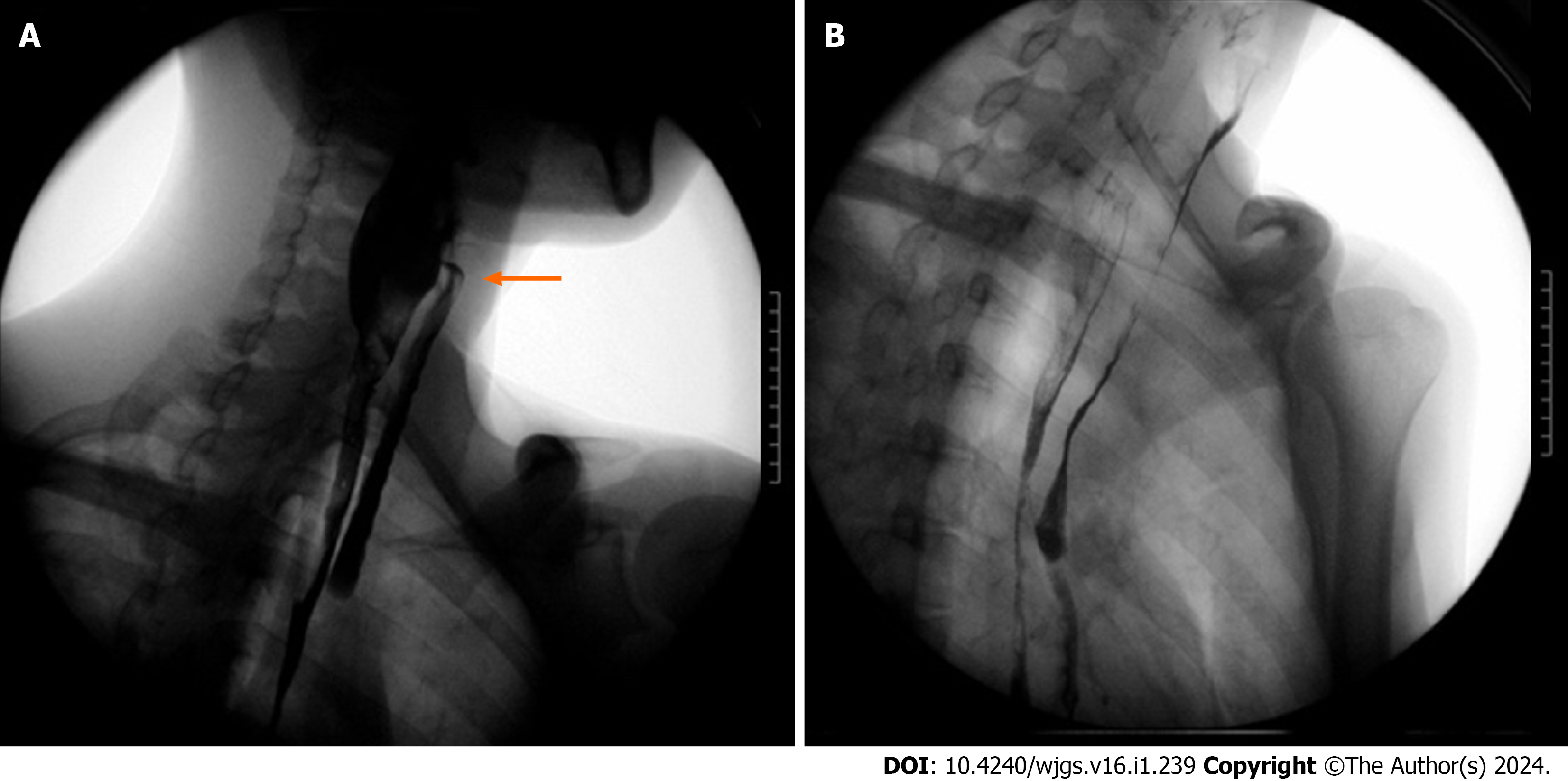
Figure 2 Upper gastrointestinal imaging showed corrosive esophageal stenosis and pharyngeal fistula.
A: Upper gastrointestinal imaging (UGI) indicated that the patient had a pharyngeal fistula (yellow arrow); B: UGI indicated that the patient had total esophageal stenosis.
- Citation: Fang JH, Li WM, He CH, Wu JL, Guo Y, Lai ZC, Li GD. Endoscopic treatment of extreme esophageal stenosis complicated with esophagotracheal fistula: A case report. World J Gastrointest Surg 2024; 16(1): 239-247
- URL: https://www.wjgnet.com/1948-9366/full/v16/i1/239.htm
- DOI: https://dx.doi.org/10.4240/wjgs.v16.i1.239


