Published online May 27, 2023. doi: 10.4240/wjgs.v15.i5.882
Peer-review started: December 12, 2022
First decision: January 2, 2023
Revised: January 16, 2023
Accepted: March 30, 2023
Article in press: March 30, 2023
Published online: May 27, 2023
Processing time: 165 Days and 7.4 Hours
Perianal fistulising Crohn's disease (PFCD) and glandular anal fistula have many similarities on conventional magnetic resonance imaging. However, many patients with PFCD show concomitant active proctitis, but only few patients with glandular anal fistula have active proctitis.
To explore the value of differential diagnosis of PFCD and glandular anal fistula by comparing the textural feature parameters of the rectum and anal canal in fat suppression T2-weighted imaging (FS-T2WI).
Patients with rectal water sac implantation were screened from the first part of this study (48 patients with PFCD and 22 patients with glandular anal fistula). Open-source software ITK-SNAP (Version 3.6.0, http://www.itksnap.org/) was used to delineate the region of interest (ROI) of the entire rectum and anal canal wall on every axial section, and then the ROIs were input in the Analysis Kit software (version V3.0.0.R, GE Healthcare) to calculate the textural feature parameters. Textural feature parameter differences of the rectum and anal canal wall between the PFCD group vs the glandular anal fistula group were analyzed using Mann-Whitney U test. The redundant textural parameters were screened by bivariate Spearman correlation analysis, and binary logistic regression analysis was used to establish the model of textural feature parameters. Finally, diagnostic accuracy was assessed by receiver operating characteristic-area under the curve (AUC) analysis.
In all, 385 textural parameters were obtained, including 37 parameters with statistically significant differences between the PFCD and glandular anal fistula groups. Then, 16 texture feature parameters remained after bivariate Spearman correlation analysis, including one histogram parameter (Histogram energy); four grey level co-occurrence matrix (GLCM) parameters (GLCM energy_all direction_offset1_SD, GLCM entropy_all direction_ offset4_SD, GLCM entropy_all direction_offset7_SD, and Haralick correlation_all direction_ offset7_SD); four texture parameters (Correlation_all direction_offset1_SD, cluster prominence _angle 90_offset4, Inertia_all direction_offset7_SD, and cluster shade_angle 45_offset7); five grey level run-length matrix parameters (grey level nonuniformity_angle 90_offset1, grey level nonuniformity_all direction_offset4_SD, long run high grey level emphasis_all direction_offset1_SD, long run emphasis_all direction_ offset4_ SD, and long run high grey level emphasis_all direction_off
The model of textural feature parameters showed good diagnostic performance for PFCD. The texture feature parameters of the rectum and anal canal in FS-T2WI are helpful to distinguish PFCD from glandular anal fistula.
Core Tip: Crohn's disease (CD) is a localized, segmental chronic granulomatous inflammation that can affect the digestive tract from the oral cavity to the anus, and its pathophysiology is non-caseous necrotic granuloma. Nearly 10% of patients with CD have an anal fistula before presenting gastrointestinal symptoms. At the same time, perianal fistulising CD (PFCD) and glandular anal fistula have many similarities on conventional magnetic resonance imaging (MRI); therefore, it is difficult to differentiate between these conditions in the early stages with conventional MRI. Texture analysis based on conventional MRI images can quantitatively analyze image pixel information and reflect the internal heterogeneity and pathological characteristics of the lesion. Currently, this approach is widely used to distinguish between benign and malignant tumors, predict tumor stage, and evaluate treatment efficacy. In addition to the application of texture analysis in the study of tumors or substantial organs, some studies have applied texture analysis to hollow organs such as the intestine. Many patients with PFCD show concomitant active proctitis, but only few patients with glandular anal fistula have active proctitis. Based on this theory, we analyzed the texture of the rectum and anal canal wall in the PFCD group and glandular anal fistula group in this study to explore whether the texture feature parameters are valuable in identifying and differentiating these two lesions.
- Citation: Zhu X, Ye DD, Wang JH, Li J, Liu SW. Diagnostic performance of texture analysis in the differential diagnosis of perianal fistulising Crohn’s disease and glandular anal fistula. World J Gastrointest Surg 2023; 15(5): 882-891
- URL: https://www.wjgnet.com/1948-9366/full/v15/i5/882.htm
- DOI: https://dx.doi.org/10.4240/wjgs.v15.i5.882
Crohn's disease (CD) is a localized, segmental chronic granulomatous inflammation that can affect the digestive tract from the oral cavity to the anus, and its pathophysiology is non-caseous necrotic granuloma[1]. Nearly 10% patients with CD have an anal fistula before presenting with gastrointestinal symptoms. At the same time, perianal fistulising CD (PFCD) and glandular anal fistula have many similarities on conventional magnetic resonance imaging (MRI); therefore, it is difficult to differentiate these conditions in the early stages with conventional MRI[2]. Texture analysis based on conventional MRI images can quantitatively analyze image pixel information and reflect the internal heterogeneity and pathological characteristics of the lesion[3]. Currently, this approach is widely used to distinguish between benign and malignant tumors, predict tumor stage, and evaluate treatment efficacy[4-7]. In addition to the application of texture analysis in the study of tumors or substantial organs, some studies have applied texture analysis to hollow organs such as the intestine[8,9]. Many patients with PFCD show concomitant active proctitis, but only few patients with glandular anal fistula have active proctitis. Based on this theory, we analyzed the texture of the rectum and anal canal wall in the PFCD group and glandular anal fistula group in this study to explore whether the texture feature parameters are valuable in identifying and differentiating these two lesions.
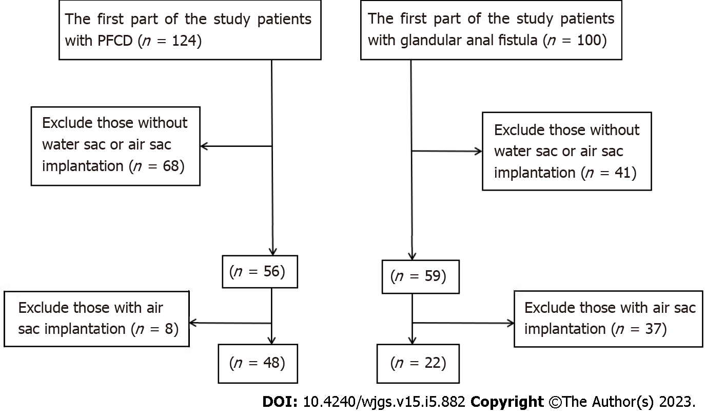
This study was approved by the institutional review board of the Affiliated Hospital of Nanjing University of Chinese Medicine; informed consent was waived owing to the retrospective nature of the study. This study was conducted in two parts through a search of our medical records. In the first part, we conducted screening for rectal water sac implantation in the existing cohort of PFCD and glandular anal fistula patients; those with an air sac or water sac were included and those without, were excluded. The flow chart of patient inclusion and exclusion is shown in Figure 1. Finally, 48 PFCD patients [41 male and 7 female; mean age: 28.60 ± 10.77 (13–61) years] with rectal water sac implantation were screened. Of these, 22 patients [19 male and 3 female; mean age: 34.95 ± 12.71 (15–62) years] had glandular anal fistula with rectal water sac implantation. This study was approved by the Ethics Review Committee of our hospital. Considering the retrospective nature of the study, the need for informed consent was waived.
Before MRI examination of the rectum, the patient was required to perform cleansing enema, and a water sac (approximately 150 mL of normal saline, fully expanded) was inserted into the rectal cavity by the clinician. All MRI exams were performed using a Siemens Magnetom Aera 1.5 T scanner, and the body phased array coil was used for scanning. The patient was in the supine position, and the scanning range was from the level of the anterior superior iliac spine to the level of the upper femur. The horizontal scanning line was perpendicular to the anal canal, while the coronal and sagittal scanning lines were parallel to the anal canal. The scanning sequence and specific parameters were the same as in the first part of the study.
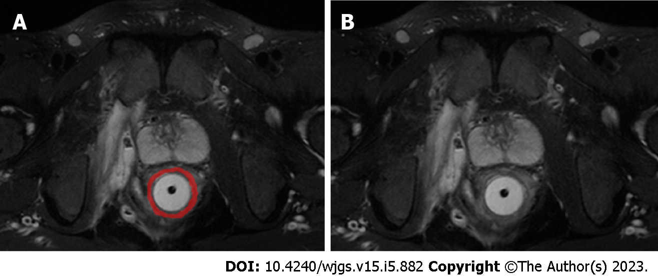
The patient’s imaging files including T2-weighted imaging (T2WI) were accessed using the open-source software ITK-SNAP (Version 3.6.0, http://www.itksnap.org/). A radiologist with 3 years diagnostic experience in anal fistula manually drew the region of interest (ROI) on the fat suppression T2WI axial position along the rectal and anal canal wall opened by the water sac (Figure 2). The scope of the sketch covered the entire rectal and anal canal wall. If there was any doubt about the sketch, another senior doctor was consulted to reach consensus. The sketched ROI and original image were imported into the Analysis Kit software (version V3.0.0.R, GE Healthcare), which automatically analyzed and calculated the texture feature parameter table of the drawn ROI.
The SPSS 22.0 software was used to perform all statistical analysis on the obtained textural feature parameters and detect the normal distribution of the parameters. For normally distributed data, two-tailed independent sample t-test was used. For non-normally distributed data, Mann–Whitney U test was used for comparison. P < 0.05 was considered to indicate statistical significance. Bivariate Spearman’s correlation analysis was conducted on indicators with statistical differences to calculate the redundancy between the textural feature parameters; those with a redundancy threshold > 0.9 were screened out. The receiver operating characteristic (ROC) curve analysis was used to calculate the diagnostic efficacy of each index selected by area under the curve (AUC) comparison, following which the Youden index, specificity, sensitivity, positive likelihood ratio, and negative likelihood ratio of each index were calculated. Finally, binary logistic regression analysis was carried out to identify indicators with statistical differences to establish the logistic regression model of textural feature parameters and calculate the corresponding statistical indicators.
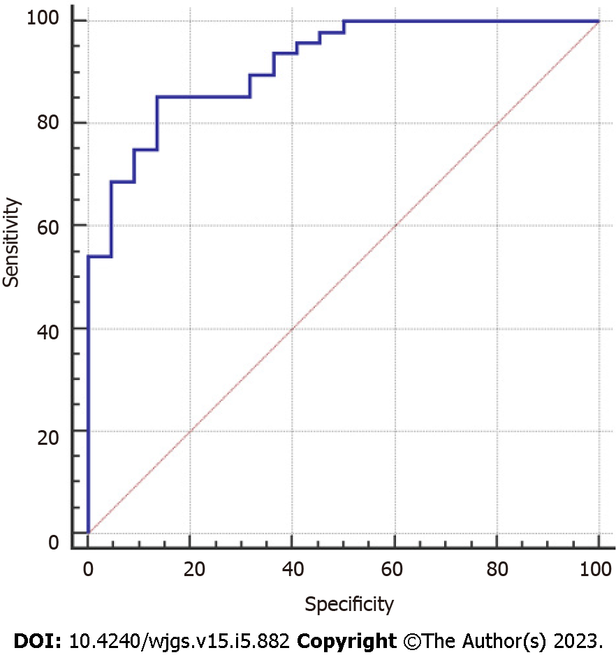
A total of 385 textural feature parameters of the final drawn ROI were calculated by the Analysis Kit software. Of these, 37 parameters showed statistically significant differences in the PFCD group and glandular anal fistula group. Through bivariate Spearman correlation analysis, the redundancy between the feature parameters was calculated, the indicators with a redundancy threshold > 0.9 were filtered out. Finally, 16 textural feature parameters were obtained, including one histogram parameter (histogram-energy); four grey level co-occurrence matrix (GLCM) parameters (GLCM energy_all direction_offset1_SD, GLCM entropy_all direction_offset4_SD, GLCM entropy_all direction_offset7_SD, and Haralick correlation_all direction_offset7_SD); four texture parameters (Correlation_all direction_offset1_SD, cluster prominence_angle 90_offset4, inertia_all direction_offset7_SD, and cluster shade_angle 45_ offset7); five grey level run-length matrix (RLM) parameters (Grey level nonuniformity_ angle 90_offset1, long run emphasis_all direction_offset4_SD, long run high grey level emphasis_all direction _offset1_SD, long run high grey level emphasis_all direction_ offset4_SD, and long run high grey level emphasis_all direction_offset4_SD); and two form factor parameters (surface area and maximum 3D diameter). ROC analysis was carried out for the above texture feature parameters. The AUC of each parameter and its corresponding Youden index, sensitivity, specificity, positive likelihood ratio, and negative likelihood ratio are shown in Table 1. Through binary logistic regression analysis, a logistic regression model of textural feature parameters was established (Table 2). The AUC of the textural feature parameter model was 0.917, and its sensitivity and specificity were 85.42% and 86.36%, respectively. The AUC, sensitivity, and specificity were higher than any individual texture feature parameters (Figure 3, Table 3).
| Textural feature parameters | AUC | Asymptotic significance | 95%CI | Z statistics | Youden index | Sensitivity | Specificity | +LR | -LR |
| Histogram parameter | |||||||||
| Histogram-energy | 0.670 | 0.0226 | 0.547-0.777 | 2.280 | 0.3068 | 62.50 | 68.18 | 1.96 | 0.55 |
| Gray-scale co-occurrence matrix parameters | |||||||||
| GLCM energy_all direction_offset1_SD | 0.683 | 0.0085 | 0.561-0.789 | 2.630 | 0.3087 | 85.42 | 45.45 | 1.57 | 0.32 |
| GLCM entropy_all direction_offset4_SD | 0.664 | 0.0185 | 0.541-0.772 | 2.354 | 0.3390 | 52.08 | 81.82 | 2.86 | 0.59 |
| GLCM entropy_all direction_ offset7_SD | 0.648 | 0.0500 | 0.524-0.758 | 1.960 | 0.2992 | 70.83 | 59.09 | 1.73 | 0.49 |
| Haralick correlation_all direction_offset7 _SD | 0.702 | 0.0027 | 0.580-0.805 | 3.005 | 0.3598 | 54.17 | 81.82 | 2.98 | 0.56 |
| Texture parameters | |||||||||
| Correlation_all direction_offset1_SD | 0.673 | 0.0126 | 0.551-0.781 | 2.496 | 0.3598 | 54.17 | 81.82 | 2.98 | 0.56 |
| Cluster prominence_angle 90_offset4 | 0.648 | 0.0300 | 0.524-0.758 | 2.171 | 0.3807 | 56.25 | 81.82 | 3.09 | 0.53 |
| Inertia_all direction_offset7_SD | 0.654 | 0.0397 | 0.531-0.764 | 2.057 | 0.3864 | 75.00 | 63.64 | 2.06 | 0.39 |
| Cluster shade_angle 45_ offset7 | 0.648 | 0.0230 | 0.524-0.758 | 2.274 | 0.3883 | 47.92 | 90.91 | 5.27 | 0.57 |
| Grey level run-length matrix (RLM) parameters | |||||||||
| Grey level nonuniformity_ angle 90_offset1 | 0.671 | 0.0144 | 0.549-0.779 | 2.446 | 0.3598 | 54.17 | 81.82 | 2.98 | 0.56 |
| Grey level nonuniformity_all direction_offset4_ SD | 0.657 | 0.0243 | 0.534-0.767 | 2.253 | 0.3125 | 81.25 | 50.00 | 0.38 | 0.62 |
| Long run high grey level emphasis_all direction _offset1_SD | 0.728 | 0.0003 | 0.609-0.828 | 3.618 | 0.3864 | 75.00 | 63.64 | 2.06 | 0.39 |
| Long run emphasis_all direction_offset4_SD | 0.722 | 0.0013 | 0.602-0.822 | 3.222 | 0.3883 | 47.92 | 90.91 | 5.27 | 0.57 |
| Long run high grey level emphasis_all direction_ offset4_SD | 0.652 | 0.0391 | 0.529-0.762 | 2.063 | 0.2803 | 91.67 | 36.36 | 1.44 | 0.23 |
| Form factor parameters | |||||||||
| Surface area | 0.728 | 0.0003 | 0.609-0.828 | 3.640 | 0.3883 | 47.92 | 90.91 | 5.27 | 0.57 |
| Maximum 3D diameter | 0.739 | 0.0001 | 0.620-0.836 | 4.024 | 0.4337 | 47.92 | 95.45 | 10.54 | 0.55 |
| β coefficient | SE | χ2 value | Significance | OR | 95%CI | ||
| Lower limit | Upper limit | ||||||
| Inertia_all direction_offset7_SD | -0.004 | 0.002 | 3.699 | 0.054 | 0.996 | 0.993 | 1.000 |
| Cluster shade_angle 45_ offset7 | 0.000 | 0.000 | 7.194 | 0.007 | 1.000 | 1.000 | 1.000 |
| Long run high grey level emphasis_all direction _offset1_SD | -0.145 | 0.069 | 4.351 | 0.037 | 0.865 | 0.755 | 0.991 |
| Long run emphasis_all direction_offset4_SD | -98.665 | 56.394 | 3.061 | 0.080 | 0.000 | 0.000 | 142353.563 |
| Surface area | 0.000 | 0.000 | 3.973 | 0.046 | 1.000 | 1.000 | 1.001 |
| Maximum 3D diameter | -0.121 | 0.040 | 8.963 | 0.003 | 0.886 | 0.819 | 0.959 |
| Constant | 9.412 | 3.758 | 6.273 | 0.012 | 12234.477 | - | - |
| AUC | Asymptotic significance level | 95%CI | Z statistics | Youden index | Sensitivity | Specificity | +LR | -LR |
| 0.917 | < 0.0001 | 0.826-0.969 | 12.645 | 0.7178 | 85.42 | 86.36 | 6.26 | 0.17 |
The textural feature parameters calculated by the Analysis Kit software were divided into the following six categories: Histogram parameters, texture parameters, form factor parameters, GLCM parameters, grey level RLM parameters, and gray-scale area matrix parameters. Among the 16 parameter indices obtained by statistical analysis in the PFCD group and the glandular anal fistula group, there were two form factor parameters, one histogram parameter, four GLCM parameters, four texture parameters, and five grey level RLM parameters.
Histogram parameters: The histogram represents the properties of a single pixel and describes the distribution of voxel intensity. The first-order statistics included 20 indicators such as energy, entropy, skewness, kurtosis, maximum intensity, minimum intensity, and mean. Among them, energy is a measure of the uniformity of the intensity level distribution, wherein a higher value represents a more uniform intensity level distribution. Entropy is a measure of the randomness of the distribution of coefficient values at intensity levels. A higher entropy value indicates more intense levels of image distribution. That is, the simpler the image, the lower the entropy value, and the more complex the image, the higher the entropy value.
Texture parameters: The texture represents the appearance of the surface and the distribution of its elements, which helps to predict the surface appearance as being either smooth or rough from the image. Correlation refers to the similarity of the gray level of adjacent pixels, indicating the correlation of pixels with their adjacent pixels in the entire image, ranging from -1 to 1. Inertia reflects the clarity and clarity of the image. The degree of depression of the texture can better distinguish the complexity of the grayscale spatial distribution of the lesion area. Cluster prominence reflects the abrupt situation of the image texture: The greater the contrast between the textures, the greater the value. Cluster shade represents the correlation between texture smoothness and symmetry: The higher the value, the less smooth and more asymmetric the texture.
Gray level co-occurrence matrix parameters: GLCM represents the joint probability of certain pixels with a certain gray value. By changing the displacement vector between each pair of pixels, the number of joint occurrences of pixels of one gray value and those of another gray value are calculated. The advantage is that it can be spatially correlated in different directions according to the spatial relationship of distance and angle, to fully display the joint information of grayscale and position. Among them, the energy represents the square sum of the elements in GLCM, the range is 0–1, the energy of the unchanged image is 1; and Haralick correlation is to measure the similarity of the gray level of the image in the row or column direction, indicating the local gray correlation. The larger the value, the greater the correlation; the sum entropy can reflect the heterogeneity of the lesion. The greater the sum entropy, the greater the qualitative nature of the disease.
Grey level run-length matrix parameters: RLM is defined as the number and running length of gray scale pixels running in a given direction. The RLM parameters reflect the roughness or smoothness of the image. The larger the long-stroke advantage value, the smoother the image. On the contrary, the larger the short-stroke advantage value, the rougher the image.
Form factor parameters: These describe the three-dimensional size and shape of the ROI[10-12].
Texture analysis is based on image images by quantifying the roughness, regularity, and uniformity of the spatial distribution of pixel gray values in normal tissues and pathological tissues to evaluate the heterogeneity of image signals (including both the heterogeneity that the human eye can and cannot recognize)[3,13]. For MRI images, the gray-scale contrast, uniformity, and texture depth and thickness of the images are important features to distinguish images of lesions from non-lesions[14]. Existing studies have used MRI texture analysis for lesion detection, classification, treatment response evaluation, and prediction of various cancers such as breast cancer, brain tumors, and rectal cancer[15-18]. The statistically different texture feature parameters obtained in this study reflect the uniformity of the distribution of PFCD and glandular anal fistula in the grayscale value of the image, correlation of local gray
Studies have found that most patients with PFCD are associated with active proctitis. Glandular anal fistula is mostly caused by infection or obstruction of the perianal glands, so it is rarely associated with active proctitis[19]. We conducted a study on the indirect signs of rectal involvement and proctitis in patients with PFCD through texture analysis of the rectal and anal canal wall in patients in the PFCD and glandular anal fistula groups. This study obtained 385 textural feature parameters from the Analysis Kit software, many of which were similar, and some features even had a negative effect on correct classification. The greater the number of features, the higher the complexity and the lower the classification speed, resulting in a reduction in classification accuracy and thus, poor universality[14]. The redundancy of each textural feature parameter was calculated by using the Spearman’s correlation analysis and finally 16 textural feature parameters were obtained. The texture feature parameter regression model was obtained by binary logistic regression. The AUC was 0.917, and the calculated sensitivity and specificity was 85.42% and 86.36%, respectively, which was higher than the AUC, sensitivity and specificity of any individual texture feature parameter. We believed that the texture feature parameters had certain discrimination value for PFCD and glandular anal fistula. Although texture analysis is rarely applied to cavity organs such as the rectum or anal canal wall, it is also applied to the analysis of the intestinal wall of Crohn's disease. Makanyanga et al[20] applied MRI texture analysis to evaluate the activity of the small intestine in CD, and found that texture feature parameters were correlated with lesion activity. Bhatnagar et al[21] found that depending on the presence or absence of histological markers of hypoxia and angiogenesis, the textural feature parameters of the T1-weighted imaging-enhanced image of the small intestine in CD were different. Both local and international literature revealed that there are no studies on the application of image texture analysis to analyze the rectal and anal canal wall of PFCD and glandular anal fistula. Our results likely show that the texture feature parameters are of certain significance for the differential diagnosis of PFCD and glandular anal fistula.
First, owing to the thin wall of the rectum and anal canal, it is difficult to delineate the ROI. In this study, filling the rectum and anal canal with water sacs was used to increase the contrast with the surrounding tissue to reduce the error. Second, because of the small sample size in this study, there was no separate verification set to validate the textural feature parameter model of this study. Further studies with larger sample size are needed to increase the stability of the textural feature parameter and verify the model. Meanwhile, the PFCD group included in this study contains a mix of patients with and without active proctitis. In the future, texture analysis should be used to further investigate whether there are differences between the two subgroups. Third, a primary issue with regard to the texture field is the decipherer of the texture features in a context, even though they were somehow validated[22].
In conclusion, the textural feature parameters obtained from the texture analysis of the rectal and anal canal wall in the PFCD group and glandular anal fistula group has some identification value for these two lesions, and can be used as a reference index for imaging specialists to identify and distinguish these two lesions.
Perianal fistulising Crohn's disease (PFCD) and glandular anal fistula have many similarities on conventional magnetic resonance imaging (MRI); therefore, it is difficult to differentiate these conditions in the early stages with conventional MRI. Texture analysis based on conventional MRI images can quantitatively analyze image pixel information and reflect the internal heterogeneity and pathological characteristics of the lesion.
This study aimed to analyze the texture of the rectum and anal canal wall in the PFCD group and glandular anal fistula group to explore whether the texture feature parameters are valuable in identifying and differentiating these two lesions, which provides a non-invasive method for preoperatively differentiating these two entities.
Therefore, the purpose of this study is to differentiate PFCD from glandular anal fistula using MRI texture analysis.
Patients with rectal water sac implantation were screened from the first part of this study (48 patients with PFCD and 22 patients with glandular anal fistula). Open-source software ITK-SNAP (Version 3.6.0, http://www.itksnap.org/) was used to delineate the region of interest (ROI) of the entire rectum and anal canal wall on every axial section, and then the ROIs were input in the Analysis Kit software (version V3.0.0.R, GE Healthcare) to calculate the textural feature parameters. Textural feature parameter differences were compared between the two groups and selected for further analysis.
In all, 385 textural parameters were obtained, including 37 parameters with statistically significant differences between the PFCD and glandular anal fistula groups. Then, 16 texture feature parameters remained after bivariate Spearman correlation analysis, including one histogram parameter; four grey level co-occurrence matrix (GLCM) parameters; four texture parameters; five grey level run-length matrix (RLM) parameters; and two form factor parameters. The AUC, sensitivity, and specificity of the model of textural feature parameters were 0.917, 85.42%, and 86.36%, respectively.
The model of textural feature parameters showed good diagnostic performance for PFCD. The texture feature parameters of the rectum and anal canal in fat suppression T2-weighted imaging are helpful to distinguish PFCD from glandular anal fistula.
This study provides a non-invasive method (MRI texture analysis) to preoperatively differentiate PFCD from glandular anal fistula, which has a profound clinical significance in guiding treatment strategy and predicting prognosis for patients with PFCD and anal fistula.
We thank our colleagues for their continuous and excellent support.
Provenance and peer review: Unsolicited article; Externally peer reviewed.
Peer-review model: Single blind
Specialty type: Gastroenterology and hepatology
Country/Territory of origin: China
Peer-review report’s scientific quality classification
Grade A (Excellent): 0
Grade B (Very good): B
Grade C (Good): C
Grade D (Fair): D
Grade E (Poor): 0
P-Reviewer: Aydin S, Turkey; Choi YS, South Korea; Garg P, India S-Editor: Li L L-Editor: A P-Editor: Wu RR
| 1. | Torres J, Mehandru S, Colombel JF, Peyrin-Biroulet L. Crohn's disease. Lancet. 2017;389:1741-1755. [RCA] [PubMed] [DOI] [Full Text] [Cited by in Crossref: 1121] [Cited by in RCA: 1788] [Article Influence: 223.5] [Reference Citation Analysis (111)] |
| 2. | Schwartz DA, Ghazi LJ, Regueiro M, Fichera A, Zoccali M, Ong EM, Mortelé KJ; Crohn's & Colitis Foundation of America, Inc. Guidelines for the multidisciplinary management of Crohn's perianal fistulas: summary statement. Inflamm Bowel Dis. 2015;21:723-730. [RCA] [PubMed] [DOI] [Full Text] [Cited by in Crossref: 52] [Cited by in RCA: 59] [Article Influence: 5.9] [Reference Citation Analysis (0)] |
| 3. | Lubner MG, Smith AD, Sandrasegaran K, Sahani DV, Pickhardt PJ. CT Texture Analysis: Definitions, Applications, Biologic Correlates, and Challenges. Radiographics. 2017;37:1483-1503. [RCA] [PubMed] [DOI] [Full Text] [Cited by in Crossref: 422] [Cited by in RCA: 571] [Article Influence: 71.4] [Reference Citation Analysis (1)] |
| 4. | Kim JH, Ko ES, Lim Y, Lee KS, Han BK, Ko EY, Hahn SY, Nam SJ. Breast Cancer Heterogeneity: MR Imaging Texture Analysis and Survival Outcomes. Radiology. 2017;282:665-675. [RCA] [PubMed] [DOI] [Full Text] [Cited by in Crossref: 151] [Cited by in RCA: 181] [Article Influence: 20.1] [Reference Citation Analysis (0)] |
| 5. | Imbriaco M, Cuocolo R. Does Texture Analysis of MR Images of Breast Tumors Help Predict Response to Treatment? Radiology. 2018;286:421-423. [RCA] [PubMed] [DOI] [Full Text] [Cited by in Crossref: 14] [Cited by in RCA: 14] [Article Influence: 2.0] [Reference Citation Analysis (0)] |
| 6. | Chamming's F, Ueno Y, Ferré R, Kao E, Jannot AS, Chong J, Omeroglu A, Mesurolle B, Reinhold C, Gallix B. Features from Computerized Texture Analysis of Breast Cancers at Pretreatment MR Imaging Are Associated with Response to Neoadjuvant Chemotherapy. Radiology. 2018;286:412-420. [RCA] [PubMed] [DOI] [Full Text] [Cited by in Crossref: 73] [Cited by in RCA: 98] [Article Influence: 12.3] [Reference Citation Analysis (0)] |
| 7. | Mulé S, Thiefin G, Costentin C, Durot C, Rahmouni A, Luciani A, Hoeffel C. Advanced Hepatocellular Carcinoma: Pretreatment Contrast-enhanced CT Texture Parameters as Predictive Biomarkers of Survival in Patients Treated with Sorafenib. Radiology. 2018;288:445-455. [RCA] [PubMed] [DOI] [Full Text] [Cited by in Crossref: 59] [Cited by in RCA: 81] [Article Influence: 11.6] [Reference Citation Analysis (0)] |
| 8. | Yin JD, Song LR, Lu HC, Zheng X. Prediction of different stages of rectal cancer: Texture analysis based on diffusion-weighted images and apparent diffusion coefficient maps. World J Gastroenterol. 2020;26:2082-2096. [RCA] [PubMed] [DOI] [Full Text] [Full Text (PDF)] [Cited by in CrossRef: 20] [Cited by in RCA: 28] [Article Influence: 5.6] [Reference Citation Analysis (0)] |
| 9. | Chen Y, Li H, Feng J, Suo S, Feng Q, Shen J. A Novel Radiomics Nomogram for the Prediction of Secondary Loss of Response to Infliximab in Crohn's Disease. J Inflamm Res. 2021;14:2731-2740. [RCA] [PubMed] [DOI] [Full Text] [Full Text (PDF)] [Cited by in Crossref: 3] [Cited by in RCA: 24] [Article Influence: 6.0] [Reference Citation Analysis (0)] |
| 10. | Yu H, Buch K, Li B, O'Brien M, Soto J, Jara H, Anderson SW. Utility of texture analysis for quantifying hepatic fibrosis on proton density MRI. J Magn Reson Imaging. 2015;42:1259-1265. [RCA] [PubMed] [DOI] [Full Text] [Cited by in Crossref: 32] [Cited by in RCA: 34] [Article Influence: 3.4] [Reference Citation Analysis (0)] |
| 11. | Li Z, Mao Y, Huang W, Li H, Zhu J, Li W, Li B. Texture-based classification of different single liver lesion based on SPAIR T2W MRI images. BMC Med Imaging. 2017;17:42. [RCA] [PubMed] [DOI] [Full Text] [Full Text (PDF)] [Cited by in Crossref: 59] [Cited by in RCA: 77] [Article Influence: 9.6] [Reference Citation Analysis (0)] |
| 12. | Chaddad A, Tanougast C. Extracted magnetic resonance texture features discriminate between phenotypes and are associated with overall survival in glioblastoma multiforme patients. Med Biol Eng Comput. 2016;54:1707-1718. [RCA] [PubMed] [DOI] [Full Text] [Cited by in Crossref: 38] [Cited by in RCA: 43] [Article Influence: 4.8] [Reference Citation Analysis (0)] |
| 13. | Sidhu HS, Benigno S, Ganeshan B, Dikaios N, Johnston EW, Allen C, Kirkham A, Groves AM, Ahmed HU, Emberton M, Taylor SA, Halligan S, Punwani S. "Textural analysis of multiparametric MRI detects transition zone prostate cancer". Eur Radiol. 2017;27:2348-2358. [RCA] [PubMed] [DOI] [Full Text] [Full Text (PDF)] [Cited by in Crossref: 69] [Cited by in RCA: 70] [Article Influence: 8.8] [Reference Citation Analysis (0)] |
| 14. | Guo CG, Ren S, Chen X, Wang QD, Xiao WB, Zhang JF, Duan SF, Wang ZQ. Pancreatic neuroendocrine tumor: prediction of the tumor grade using magnetic resonance imaging findings and texture analysis with 3-T magnetic resonance. Cancer Manag Res. 2019;11:1933-1944. [RCA] [PubMed] [DOI] [Full Text] [Full Text (PDF)] [Cited by in Crossref: 47] [Cited by in RCA: 48] [Article Influence: 8.0] [Reference Citation Analysis (0)] |
| 15. | Su CQ, Lu SS, Han QY, Zhou MD, Hong XN. Intergrating conventional MRI, texture analysis of dynamic contrast-enhanced MRI, and susceptibility weighted imaging for glioma grading. Acta Radiol. 2019;60:777-787. [RCA] [PubMed] [DOI] [Full Text] [Cited by in Crossref: 13] [Cited by in RCA: 26] [Article Influence: 4.3] [Reference Citation Analysis (0)] |
| 16. | Parikh J, Selmi M, Charles-Edwards G, Glendenning J, Ganeshan B, Verma H, Mansi J, Harries M, Tutt A, Goh V. Changes in primary breast cancer heterogeneity may augment midtreatment MR imaging assessment of response to neoadjuvant chemotherapy. Radiology. 2014;272:100-112. [RCA] [PubMed] [DOI] [Full Text] [Cited by in Crossref: 109] [Cited by in RCA: 108] [Article Influence: 9.8] [Reference Citation Analysis (0)] |
| 17. | Meng Y, Zhang C, Zou S, Zhao X, Xu K, Zhang H, Zhou C. MRI texture analysis in predicting treatment response to neoadjuvant chemoradiotherapy in rectal cancer. Oncotarget. 2018;9:11999-12008. [RCA] [PubMed] [DOI] [Full Text] [Full Text (PDF)] [Cited by in Crossref: 38] [Cited by in RCA: 30] [Article Influence: 4.3] [Reference Citation Analysis (0)] |
| 18. | De Cecco CN, Ganeshan B, Ciolina M, Rengo M, Meinel FG, Musio D, De Felice F, Raffetto N, Tombolini V, Laghi A. Texture analysis as imaging biomarker of tumoral response to neoadjuvant chemoradiotherapy in rectal cancer patients studied with 3-T magnetic resonance. Invest Radiol. 2015;50:239-245. [RCA] [PubMed] [DOI] [Full Text] [Cited by in Crossref: 145] [Cited by in RCA: 159] [Article Influence: 15.9] [Reference Citation Analysis (0)] |
| 19. | Tougeron D, Savoye G, Savoye-Collet C, Koning E, Michot F, Lerebours E. Predicting factors of fistula healing and clinical remission after infliximab-based combined therapy for perianal fistulizing Crohn's disease. Dig Dis Sci. 2009;54:1746-1752. [RCA] [PubMed] [DOI] [Full Text] [Cited by in Crossref: 63] [Cited by in RCA: 52] [Article Influence: 3.3] [Reference Citation Analysis (0)] |
| 20. | Makanyanga J, Ganeshan B, Rodriguez-Justo M, Bhatnagar G, Groves A, Halligan S, Miles K, Taylor SA. MRI texture analysis (MRTA) of T2-weighted images in Crohn's disease may provide information on histological and MRI disease activity in patients undergoing ileal resection. Eur Radiol. 2017;27:589-597. [RCA] [PubMed] [DOI] [Full Text] [Full Text (PDF)] [Cited by in Crossref: 24] [Cited by in RCA: 35] [Article Influence: 3.9] [Reference Citation Analysis (0)] |
| 21. | Bhatnagar G, Makanyanga J, Ganeshan B, Groves A, Rodriguez-Justo M, Halligan S, Taylor SA. MRI texture analysis parameters of contrast-enhanced T1-weighted images of Crohn's disease differ according to the presence or absence of histological markers of hypoxia and angiogenesis. Abdom Radiol (NY). 2016;41:1261-1269. [RCA] [PubMed] [DOI] [Full Text] [Full Text (PDF)] [Cited by in Crossref: 9] [Cited by in RCA: 16] [Article Influence: 1.8] [Reference Citation Analysis (0)] |
| 22. | Ren S, Zhao R, Zhang J, Guo K, Gu X, Duan S, Wang Z, Chen R. Diagnostic accuracy of unenhanced CT texture analysis to differentiate mass-forming pancreatitis from pancreatic ductal adenocarcinoma. Abdom Radiol (NY). 2020;45:1524-1533. [RCA] [PubMed] [DOI] [Full Text] [Cited by in Crossref: 27] [Cited by in RCA: 25] [Article Influence: 5.0] [Reference Citation Analysis (0)] |


