Copyright
©The Author(s) 2023.
World J Gastrointest Surg. Apr 27, 2023; 15(4): 592-599
Published online Apr 27, 2023. doi: 10.4240/wjgs.v15.i4.592
Published online Apr 27, 2023. doi: 10.4240/wjgs.v15.i4.592
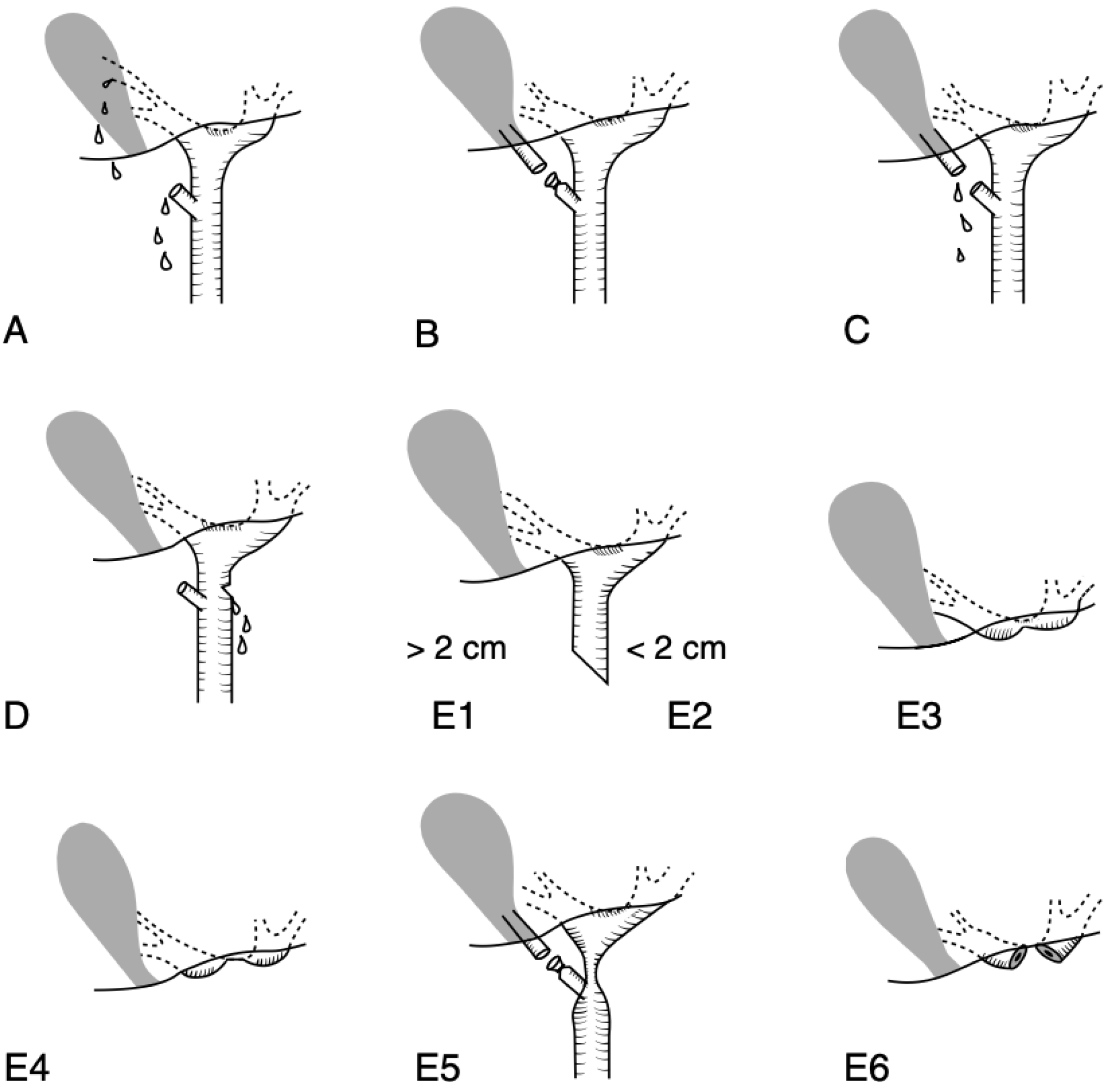
Figure 1 Classification of the bile duct injuries after cholecystectomy.
A: Schematic classification of bile duct injuries in laparoscopic cholecystectomy. Originally developed by Strasberg et al in 1995[7]. A bile leak from the cystic duct stump or minor biliary radical in the gallbladder fossa; B: Occluded right posterior sectoral duct; C: Bile leak from the divided right posterior sectoral duct; D: Bile leak from the main bile duct without major tissue loss; E1: Transected main bile duct with a stricture more than 2 cm from the hilus; E2: Transected main bile duct with a stricture less than 2 cm from the hilus; E3: Stricture of the hilus with right and left ducts in communication; E4: Stricture of the hilus with separation of right and left ducts; E5: Stricture of the main bile duct and the right posterior sectoral duct; E6: complete excision of the extrahepatic ducts involving the confluence (this injury is not described in Strasberg’s original classification). Citation: Connor S, Garden OJ. Bile duct injury in the era of laparoscopic cholecystectomy. Br J Surg 2006; 93(2): 158-168. Copyright ©John Wiley & Son´s Ltd 2006. Published by John Wiley & Son´s Ltd[8].
- Citation: Siiki A, Ahola R, Vaalavuo Y, Antila A, Laukkarinen J. Initial management of suspected biliary injury after laparoscopic cholecystectomy. World J Gastrointest Surg 2023; 15(4): 592-599
- URL: https://www.wjgnet.com/1948-9366/full/v15/i4/592.htm
- DOI: https://dx.doi.org/10.4240/wjgs.v15.i4.592


