Published online Oct 27, 2023. doi: 10.4240/wjgs.v15.i10.2351
Peer-review started: June 5, 2023
First decision: July 23, 2023
Revised: August 4, 2023
Accepted: August 27, 2023
Article in press: August 27, 2023
Published online: October 27, 2023
Processing time: 144 Days and 8.3 Hours
Few reports have described living foreign bodies in the human body. The current manuscript demonstrates that computed tomography (CT) is an effective tool for accurate preoperative evaluation of living foreign bodies in clinic. The three-dimensional (3D) reconstruction technology could clearly display anatomical stru
Herein we describe a 68-year-old man diagnosed with digestive tract perforation and acute peritonitis caused by a foreign body of Monopterus albus. The patient pre
The current manuscript demonstrates that CT is an effective tool for accurate preoperative evaluation of living foreign bodies in clinic.
Core Tip: Computed tomography (CT) is an effective tool for accurate preoperative evaluation of living foreign bodies in the human body. Three-dimensional (3D) CT reconstruction could clearly display anatomical structures, lesions and adjacent organs, improving diagnostic accuracy. In the present case, preoperative 3D CT reconstruction accurately showed a foreign body located outside the intestinal cavity with a perforation site, and revealed that the foreign body had damaged the mesentery in the small intestine, causing fluid and gas accumulation, as well as peritoneal thickening. These findings indicate preoperative 3D CT reconstruction may accurately locate perforation sites and foreign bodies, help diagnose peri
- Citation: Yang JH, Lan JY, Lin AY, Huang WB, Liao JY. Three-dimensional computed tomography reconstruction diagnosed digestive tract perforation and acute peritonitis caused by Monopterus albus: A case report. World J Gastrointest Surg 2023; 15(10): 2351-2356
- URL: https://www.wjgnet.com/1948-9366/full/v15/i10/2351.htm
- DOI: https://dx.doi.org/10.4240/wjgs.v15.i10.2351
Digestive tract perforation is a common acute abdominal pathology[1,2], often secondary to ulcers, trauma, inflammation, tumors, etc. Computed tomography (CT) constitutes an effective tool for accurate preoperative evaluation of foreign bodies in clinic[3]. Preoperative three-dimensional (3D) CT reconstruction accurately locates perforation sites and foreign bodies, helps diagnose peritonitis and guides surgical treatment[4]. In the present case, according to clinical symptoms and signs, combined with plain 3D CT reconstruction, it was determined that the patient had digestive tract perforation, and a Monopterus albus had died after entering the abdominal cavity[5]. As a result, the patient’s abdominal cavity was seriously polluted, with a large amount of turbid yellow fluid and a small amount of feces attached to several intestinal areas, so it could be determined that the patient had “intestinal perforation” caused by a Monopterus albus[6]. The intes
One patient, a 68-year-old man from China, presented to the hospital’s emergency department after suffering from dull abdominal pain, profuse sweating and a pale complexion during the two-hour workday.
Symptoms started 2 h before presentation with complaints of dull abdominal pain, profuse sweating and a pale com
The patient didn’t have any remarkable history.
The patient denied having a family history of any malignant tumors.
Using a physical examination, the results showed the following vital signs: Blood pressure, 118/69 mmHg; body tem
Laboratory tests showed normal liver function, alpha-fetoprotein, carbohydrate antigen 19-9, and carcinoembryonic antigen. No abnormality was found in routine blood and urine analyses. Primary laboratory data upon admission are summarized in Table 1.
| Parameter | Value (admission) | Reference value | Unit |
| N-terminal-pro B-type natriuretic peptide | 80 | 0-125 | pg/mL |
| Urea | 3.7 | 2.5-6.1 | mmol/L |
| Creatinine | 58 | 46-92 | μmol/L |
| Troponin | < 0.01 | 0-0.04 | ng/mL |
| Myoglobin | 111.1 | 0-120 | ng/mL |
| Carcinoembryonic antigen | 4.5 | 0-5.0 | ng/mL |
| Carbohydrate antigen 19-9 | < 3 | 0-27 | U/mL |
| Alpha-fetoprotein | 3.2 | 0-7.000 | ng/mL |
| Creatine kinase | 102.2 | 30-135 | U/L |
| Creatine kinase MB isoenzyme | 11.3 | 0-16 | U/L |
| Lactic dehydrogenase | 145.88 | 120-246 | U/L |
| D-dimer | 0.28 | 0-0.5 | μg/mL |
| White blood cell | 9.10 | 4-10 | 109/L |
| Platelet | 140 | 125-350 | 109/L |
| Hemoglobin | 119 | 110-150 | g/L |
| Alanine aminotransferase | 18.28 | 5-40 | IU/L |
| Aspartate aminotransferase | 15.44 | 8-40 | IU/L |
| Total bilirubin | 16.48 | 5-21 | μmol/L |
| Direct bilirubin | 3.26 | 0-3.4 | μmol/L |
| Indirect bilirubin | 13.12 | 1.6-21 | μmol/L |
| Albumin | 42.73 | 35-52 | g/L |
| Total cholesterol | 3.67 | 3.0-5.7 | mmol/L |
| Low density lipoprotein cholesterol | 3.65 | < 4.13 | mmol/L |
| High density lipoprotein cholesterol | 1.86 | 1.29-1.55 | mmol/L |
| Fasting blood glucose | 4.44 | 3.9-6.1 | Mmol/L |
| Urinary protein | negative | negative | - |
| Hepatitis B surface antigen | 0 | < 0.05 | IU/mL |
| Antibody to hepatitis C | 6.04 | < 1 | S/CO |
| Immunodeficiency virus antigen and antibody | 0.09 | < 1 | S/CO |
| Antibody to treponema pallidum | 0.07 | < 1 | S/CO |
| Hepatitis C virus RNA | 0 | 0 | IU/mL |
| Anti-nuclear antibodies | negative | negative | - |
| Anti-cyclic citrullinated peptide antibody | 13.89 | < 25 | RU/mL |
| Anti-cardiolipin antibody | 2.23 | 0-12 | RU/mL |
| Blood ammonia | 22 | 9-30 | μmol/L |
| Erythrocyte sedimentation rate | 15 | 0-20 | mm/h |
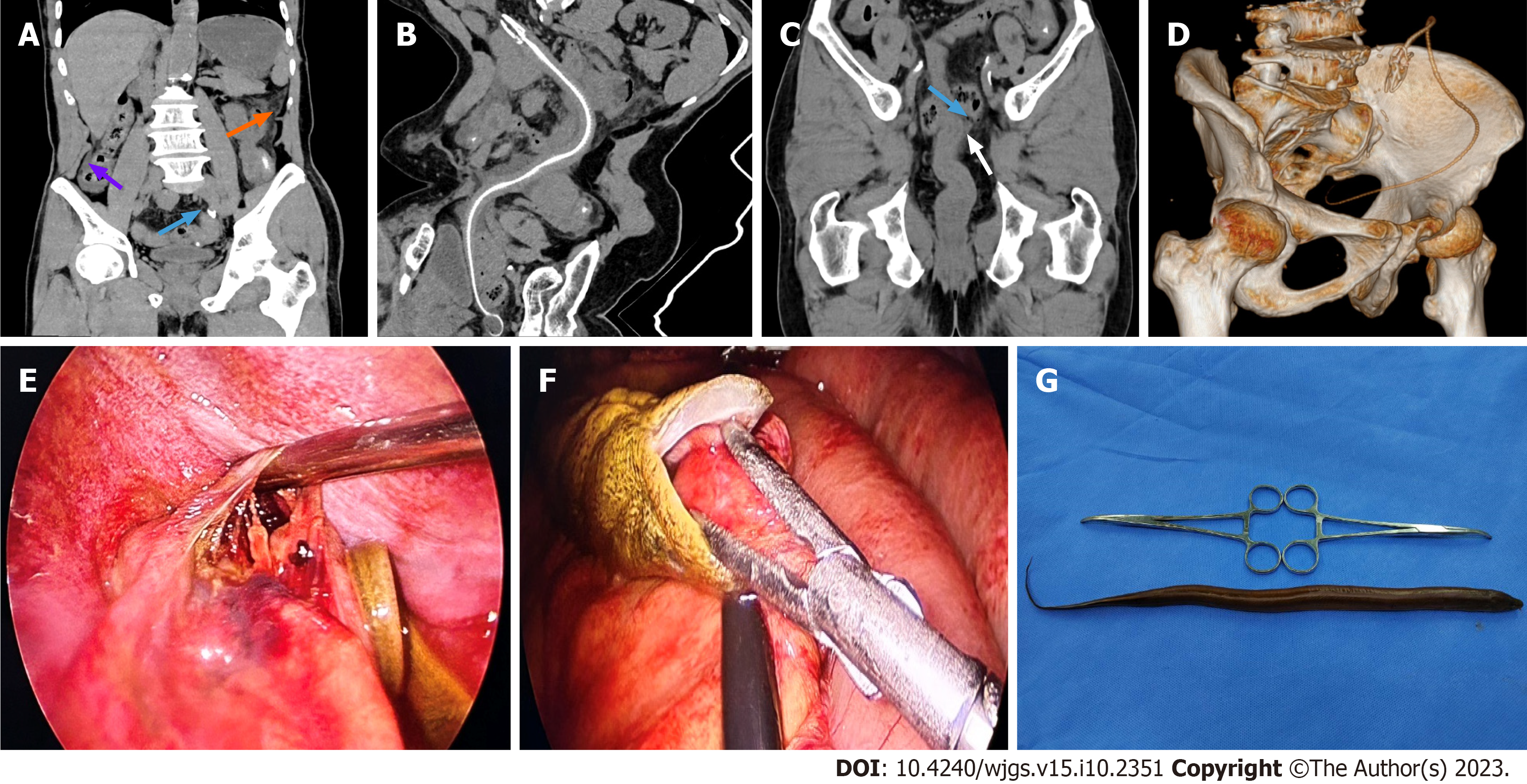
CT with multi-plane reconstruction revealed scattered exudation, effusion and free gas in the abdominal cavity, indi
The patient consented to laparoscopic surgery. Laparoscopic exploration revealed abundant cloudy yellow fluid and small amounts of feces-like fluid in the abdominal and pelvic cavities, and a large perforation was detected in the mid-rectum, with a diameter approximating 1.5 cm (Figure 1E), alongside a small amount of stool. It could be observed that the Monopterus albus has completely entered the abdominal cavity and has tightly bitten the mesentery of the small intes
Based on the patient’s previous medical history, the patient was eventually diagnosed with digestive tract perforation and acute peritonitis.
Postoperatively, the patient recovered well and was discharged on postoperative 5 d.
The patient recovered without complications.
Few reports have described living foreign bodies in the human body[9]. CT constitutes an effective tool for accurate preoperative evaluation of living foreign bodies in clinic[3]. 3D CT reconstruction clearly displays anatomical structures, lesions and adjacent organs, improving diagnostic accuracy[6]. In the present case, preoperative 3D CT reconstruction accurately located a Monopterus albus outside the intestinal cavity with a perforation site, and the foreign body had damaged the mesentery in the small intestine, causing fluid and gas accumulation, as well as peritoneal thickening[5]. These findings suggest preoperative 3D CT reconstruction may accurately locate perforation sites and foreign bodies, help diagnose peritonitis and guide surgical treatment[4].
Preoperative 3D CT reconstruction can accurately locate perforation sites and living foreign bodies, help diagnose peritonitis and guide surgical treatment.
Provenance and peer review: Unsolicited article; Externally peer reviewed.
Peer-review model: Single blind
Specialty type: Gastroenterology and hepatology
Country/Territory of origin: China
Peer-review report’s scientific quality classification
Grade A (Excellent): 0
Grade B (Very good): B, B
Grade C (Good): 0
Grade D (Fair): 0
Grade E (Poor): 0
P-Reviewer: Kumar M, India; Oley MH, Indonesia S-Editor: Wang JJ L-Editor: A P-Editor: Wang JJ
| 1. | Chen P, Gao J, Li J, Yu R, Wang L, Xue F, Zheng X, Gao L, Shang X. Construction and efficacy evaluation of an early warning scoring system for septic shock in patients with digestive tract perforation: A retrospective cohort study. Front Med (Lausanne). 2022;9:976963. [RCA] [PubMed] [DOI] [Full Text] [Full Text (PDF)] [Reference Citation Analysis (3)] |
| 2. | Hajibandeh S, Shah J, Hajibandeh S, Murali S, Stephanos M, Ibrahim S, Asqalan A, Mithany R, Wickramasekara N, Mansour M. Intraperitoneal contamination index (Hajibandeh index) predicts nature of peritoneal contamination and risk of postoperative mortality in patients with acute abdominal pathology: a prospective multicentre cohort study. Int J Colorectal Dis. 2021;36:1023-1031. [RCA] [PubMed] [DOI] [Full Text] [Cited by in Crossref: 2] [Cited by in RCA: 6] [Article Influence: 1.5] [Reference Citation Analysis (0)] |
| 3. | Kang J, Jiang L. Application Value of the CT Scan 3D Reconstruction Technique in Maxillofacial Fracture Patients. Evid Based Complement Alternat Med. 2022;2022:1643434. [RCA] [PubMed] [DOI] [Full Text] [Full Text (PDF)] [Reference Citation Analysis (0)] |
| 4. | Lee CM, Liu RW. Comparison of pelvic incidence measurement using lateral x-ray, standard ct versus ct with 3d reconstruction. Eur Spine J. 2022;31:241-247. [RCA] [PubMed] [DOI] [Full Text] [Cited by in RCA: 12] [Reference Citation Analysis (0)] |
| 5. | Kathayat LB, Chalise A, Maharjan JS, Bajracharya J, Shrestha R. Intestinal Perforation with Ingestion of Blunt Foreign Bodies: A Case Report. JNMA J Nepal Med Assoc. 2022;60:817-820. [RCA] [PubMed] [DOI] [Full Text] [Cited by in Crossref: 2] [Cited by in RCA: 2] [Article Influence: 0.7] [Reference Citation Analysis (0)] |
| 6. | Shinde RK, Rajendran R Jr. Spontaneous Intestinal Perforation in Neonates Involving the Cecum: A Case Report. Cureus. 2023;15:e37322. [RCA] [PubMed] [DOI] [Full Text] [Reference Citation Analysis (0)] |
| 7. | Hoshino N, Endo H, Hida K, Kumamaru H, Hasegawa H, Ishigame T, Kitagawa Y, Kakeji Y, Miyata H, Sakai Y. Laparoscopic Surgery for Acute Diffuse Peritonitis Due to Gastrointestinal Perforation: A Nationwide Epidemiologic Study Using the National Clinical Database. Ann Gastroenterol Surg. 2022;6:430-444. [RCA] [PubMed] [DOI] [Full Text] [Full Text (PDF)] [Cited by in RCA: 12] [Reference Citation Analysis (0)] |
| 8. | Lee MO, Jin SY, Lee SK, Hwang S, Kim TG, Song YG. Video-assisted thoracoscopic surgical wedge resection using multiplanar computed tomography reconstruction-fluoroscopy after CT guided microcoil localization. Thorac Cancer. 2021;12:1721-1725. [RCA] [PubMed] [DOI] [Full Text] [Full Text (PDF)] [Cited by in Crossref: 1] [Cited by in RCA: 5] [Article Influence: 1.3] [Reference Citation Analysis (0)] |
| 9. | Withers BT. Foreign bodies of the larynx; with report of an unusual case. Ann Otol Rhinol Laryngol. 1949;58:1085-1092. [RCA] [PubMed] [DOI] [Full Text] [Cited by in Crossref: 2] [Cited by in RCA: 2] [Article Influence: 0.0] [Reference Citation Analysis (0)] |


