Copyright
©The Author(s) 2017.
World J Gastrointest Surg. Aug 27, 2017; 9(8): 174-181
Published online Aug 27, 2017. doi: 10.4240/wjgs.v9.i8.174
Published online Aug 27, 2017. doi: 10.4240/wjgs.v9.i8.174
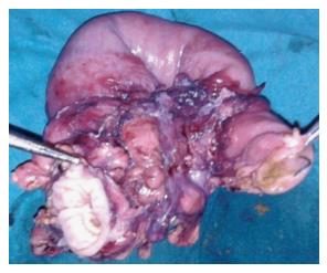
Figure 1 Intestinal tuberculosis (ileocaecal) (with permission from Chumber et al[51], 2001).
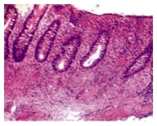
Figure 2 Histopathology (H/E stain): Showing multiple mucosal and submucosal epitheloid cell granulomas with Langhan’s giant cells in a case of colonic tuberculosis (with permission from Tandon et al[35], 1972).
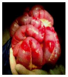
Figure 3 Peritoneal tuberculosis (with permission from Bolognesi et al[43], 2013).
- Citation: Weledji EP, Pokam BT. Abdominal tuberculosis: Is there a role for surgery? World J Gastrointest Surg 2017; 9(8): 174-181
- URL: https://www.wjgnet.com/1948-9366/full/v9/i8/174.htm
- DOI: https://dx.doi.org/10.4240/wjgs.v9.i8.174


