Copyright
©The Author(s) 2017.
World J Gastrointest Surg. Mar 27, 2017; 9(3): 92-96
Published online Mar 27, 2017. doi: 10.4240/wjgs.v9.i3.92
Published online Mar 27, 2017. doi: 10.4240/wjgs.v9.i3.92
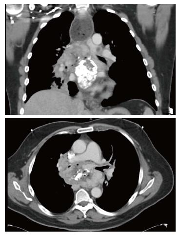
Figure 1 Coronal and axial computed tomography view of the partially calcified subcarinal mass surrounding the carina, the bronchi, and eroding into the esophagus (note the mediastinal air).
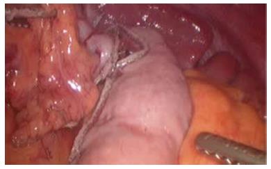
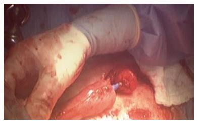
Figure 2 Intraoperative view of the gastric conduit after complete tubularization.
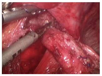
Figure 3 Dissection of the esophageal stump into the mediastinum.
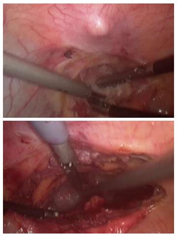
Figure 4 A wide substernal tunnel is created under direct visualization immediately posterior to the xyphoid process.
Figure 5 Circular mechanical stapled anastomosis at the neck.
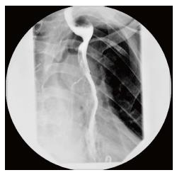
Figure 6 Esophagram demonstrating normal transit of contrast trough the anastomosis and prompt gastric emptying.
- Citation: Mungo B, Barbetta A, Lidor AO, Stem M, Molena D. Laparoscopic retrosternal gastric pull-up for fistulized mediastinal mass. World J Gastrointest Surg 2017; 9(3): 92-96
- URL: https://www.wjgnet.com/1948-9366/full/v9/i3/92.htm
- DOI: https://dx.doi.org/10.4240/wjgs.v9.i3.92


