Copyright
©The Author(s) 2017.
World J Gastrointest Surg. Oct 27, 2017; 9(10): 209-213
Published online Oct 27, 2017. doi: 10.4240/wjgs.v9.i10.209
Published online Oct 27, 2017. doi: 10.4240/wjgs.v9.i10.209
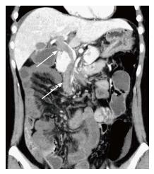
Figure 1 Abdominal computed tomography image obtained at the initial examination.
Acute mesenteric vein thrombosis extending into the portal vein (arrow) was demonstrated.
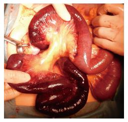
Figure 2 Gangrenous portion of the small intestine.
A gangrenous portion of the small intestine extending from 80 cm distal to the ligament of Treitz to 160 cm proximal to the ileocecal valve was found.
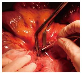
Figure 3 Surgical removal of superior mesenteric vein thrombi with a Fogarty catheter.
A Fogarty catheter was inserted from superior mesenteric vein proximal to the ileocolic vein (arrow). The thrombus was removed and blood flow was confirmed.
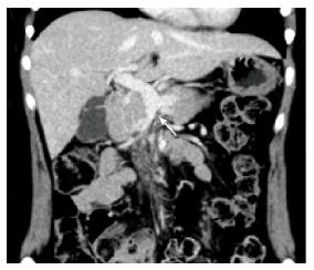
Figure 4 Abdominal computed tomography image obtained four months after surgery.
The portal vein recanalized completely, and the superior mesenteric vein was completely occluded from the distal to the first jejunal branches (arrow).
- Citation: Hirata M, Yano H, Taji T, Shirakata Y. Mesenteric vein thrombosis following impregnation via in vitro fertilization-embryo transfer. World J Gastrointest Surg 2017; 9(10): 209-213
- URL: https://www.wjgnet.com/1948-9366/full/v9/i10/209.htm
- DOI: https://dx.doi.org/10.4240/wjgs.v9.i10.209


