Copyright
©The Author(s) 2017.
World J Gastrointest Surg. Jan 27, 2017; 9(1): 1-12
Published online Jan 27, 2017. doi: 10.4240/wjgs.v9.i1.1
Published online Jan 27, 2017. doi: 10.4240/wjgs.v9.i1.1
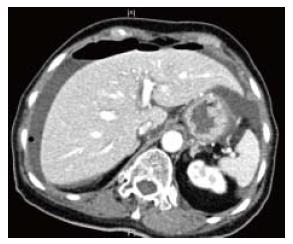
Figure 1 Computerized tomography scan shows free air under the diaphragm with peri-hepatic free fluid.
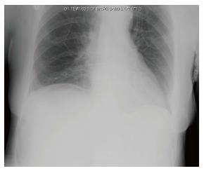
Figure 2 Erect chest X-ray image of the same patient with equivocal free air under the right hemidiaphragm.
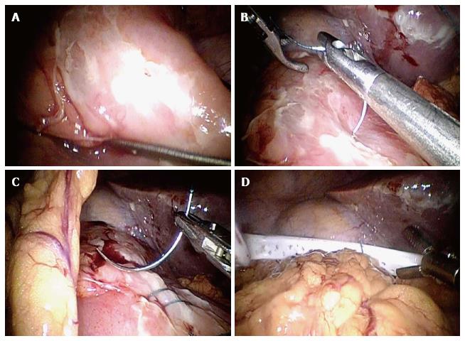
Figure 3 Shows laparoscopic omental patch repair.
A: Anterior duodenal perforation; B: Laparoscopic suturing; C: Omental patch; D: Abdominal drain placement.
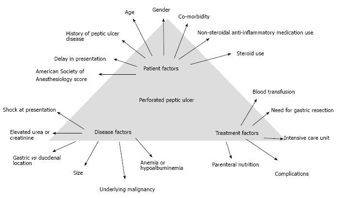
Figure 4 Determinants of outcomes in patients with perforated peptic ulcer.
- Citation: Chung KT, Shelat VG. Perforated peptic ulcer - an update. World J Gastrointest Surg 2017; 9(1): 1-12
- URL: https://www.wjgnet.com/1948-9366/full/v9/i1/1.htm
- DOI: https://dx.doi.org/10.4240/wjgs.v9.i1.1


