Copyright
©The Author(s) 2016.
World J Gastrointest Surg. Sep 27, 2016; 8(9): 651-655
Published online Sep 27, 2016. doi: 10.4240/wjgs.v8.i9.651
Published online Sep 27, 2016. doi: 10.4240/wjgs.v8.i9.651
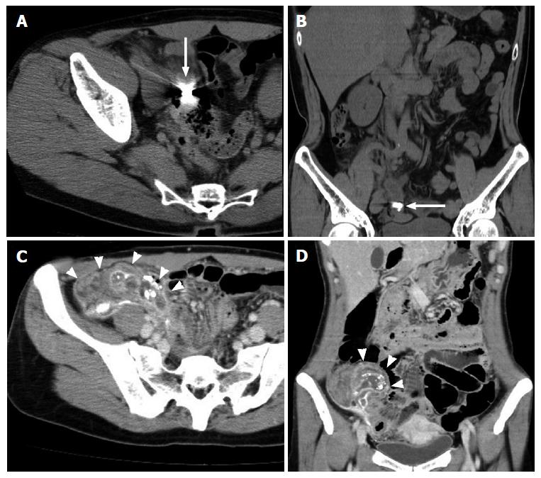
Figure 1 Abdominal computed tomography scans with axial and coronal views.
A and B: High density material is seen inside the swollen appendix (arrows); C and D: High density material is seen inside the swollen appendix and in the peritoneal cavity with a fluid collection (arrow heads). This strongly suggests a perforated appendicitis with residual barium.
- Citation: Katagiri H, Lefor AK, Kubota T, Mizokami K. Barium appendicitis: A single institution review in Japan. World J Gastrointest Surg 2016; 8(9): 651-655
- URL: https://www.wjgnet.com/1948-9366/full/v8/i9/651.htm
- DOI: https://dx.doi.org/10.4240/wjgs.v8.i9.651


