Copyright
©The Author(s) 2016.
World J Gastrointest Surg. Jul 27, 2016; 8(7): 513-520
Published online Jul 27, 2016. doi: 10.4240/wjgs.v8.i7.513
Published online Jul 27, 2016. doi: 10.4240/wjgs.v8.i7.513
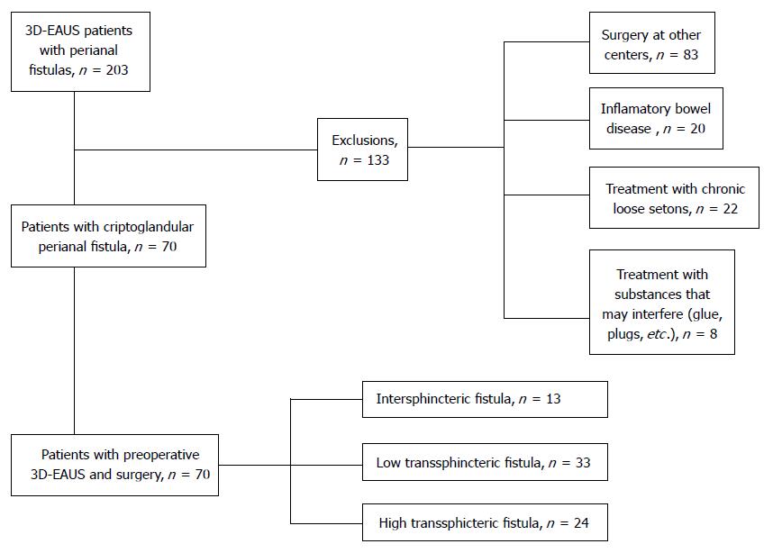
Figure 1 Patient distribution.
3D-EAUS: Three-dimensional endoanal ultrasound.
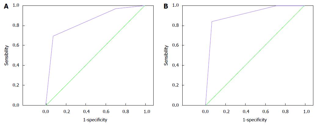
Figure 2 Receiver operating characteristic curves for the diagnosis of high transsphincteric fistulas with two-dimensional endoanal ultrasound (A) (area under curve = 0.
842; 95%CI: 0.745-0.939; P = 0.0001) and three-dimensional endoanal ultrasound (B) (area under curve = 0.910; 95%CI: 0.835-0.985; P = 0.0001).
- Citation: Garcés-Albir M, García-Botello SA, Espi A, Pla-Martí V, Martin-Arevalo J, Moro-Valdezate D, Ortega J. Three-dimensional endoanal ultrasound for diagnosis of perianal fistulas: Reliable and objective technique. World J Gastrointest Surg 2016; 8(7): 513-520
- URL: https://www.wjgnet.com/1948-9366/full/v8/i7/513.htm
- DOI: https://dx.doi.org/10.4240/wjgs.v8.i7.513


