Copyright
©The Author(s) 2016.
World J Gastrointest Surg. Mar 27, 2016; 8(3): 222-231
Published online Mar 27, 2016. doi: 10.4240/wjgs.v8.i3.222
Published online Mar 27, 2016. doi: 10.4240/wjgs.v8.i3.222
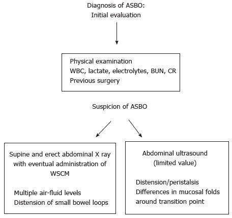
Figure 1 Adhesive small bowel obstruction diagnosis: Initial evaluation.
ASBO: Adhesive small bowel obstruction; WBC: White blood cell count; BUN: Blood urea nitrogen; CR: Creatinine; WSCM: Water soluble contrast medium.
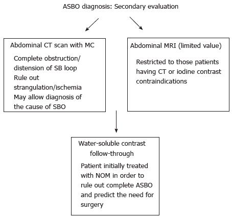
Figure 2 Adhesive small bowel obstruction diagnosis: Secondary evaluation.
ASBO: Adhesive small bowel obstruction; NOM: Non operative management; CT: Computed tomography; MC: Medium contrast.
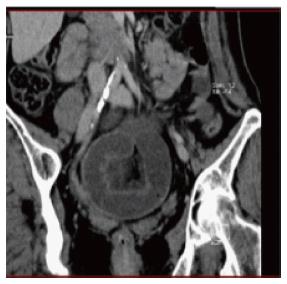
Figure 3 Adhesive small bowel obstruction caused by single band adhesion: Computed tomography scan.
Figure 4 Adhesive small bowel obstruction treatment.
ASBO: Adhesive small bowel obstruction; NGT: Naso-gastric tube; LT: Long tube.
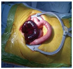
Figure 5 Adhesive small bowel obstruction caused by single band adhesion: Open surgery.
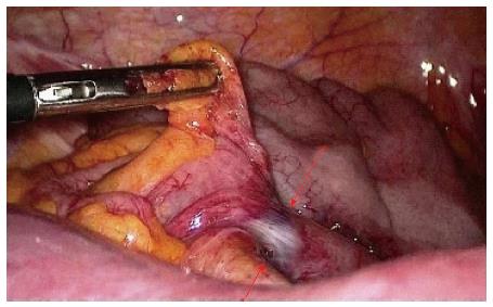
Figure 6 Adhesive small bowel obstruction caused by single band adhesion: Laparoscopic surgery.
- Citation: Catena F, Di Saverio S, Coccolini F, Ansaloni L, De Simone B, Sartelli M, Van Goor H. Adhesive small bowel adhesions obstruction: Evolutions in diagnosis, management and prevention. World J Gastrointest Surg 2016; 8(3): 222-231
- URL: https://www.wjgnet.com/1948-9366/full/v8/i3/222.htm
- DOI: https://dx.doi.org/10.4240/wjgs.v8.i3.222


