Copyright
©The Author(s) 2016.
World J Gastrointest Surg. Mar 27, 2016; 8(3): 202-211
Published online Mar 27, 2016. doi: 10.4240/wjgs.v8.i3.202
Published online Mar 27, 2016. doi: 10.4240/wjgs.v8.i3.202
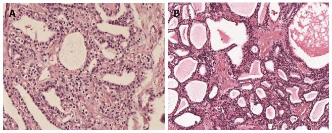
Figure 1 Pathological examinations revealing that serous cystic neoplasm cystic walls are lined with cubic flat epithelia consisting of glycogen-rich, watery-fluid-producing cells (hematoxylin and eosin × 100 ).
A: Pathology of a microcystic SCN of the pancreas; B: Pathology of a macrocystic SCN of the pancreas. SCN: Serous cystic neoplasm.
Figure 2 Microcystic pancreatic serous cystic neoplasm presentation on computed tomography/magnetic resonance imaging.
A: A microcystic pancreatic SCN lesion was revealed in the tail of the pancreas; B: MRI showed a microcystic lesion in the body of pancreas; C: A microcystic SCN lesion was revealed by magnetic resonance cholangiopancreatography. Images B and C came from the same patient. SCN: Serous cystic neoplasm; MRI: Magnetic resonance imaging.
Figure 3 Solid variant microcystic serous cystic neoplasm with honeycomb characteristics.
A: Gross pathology of a solid variant microcystic SCN; B: Histology of solid variant microcystic SCN; C: Solid variant lesion was detected by CT; D: Solid variant lesion was detected by contrast-enhanced CT. These four images were acquired from the same patient. SCN: Serous cystic neoplasm; CT: Computed tomography.
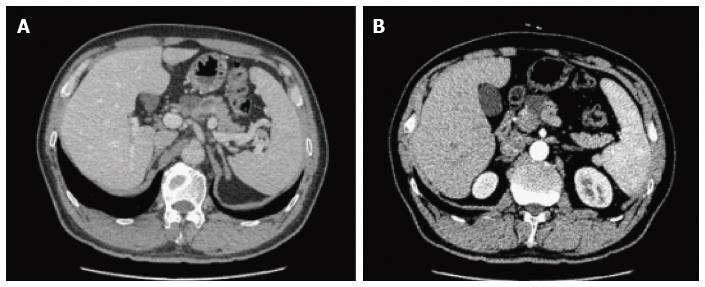
Figure 4 A macrocystic pancreatic serous cystic neoplasm was detected on computed tomography with (B) and without enhancement (A).
Figure 5 A microcystic pancreatic serous cystic neoplasm was discovered by endoscopic ultrasonography.
Figure 6 Central calcifications were found by computed tomography with and without enhancement (A and B), and also shown after three dimensional reconstruction (C).
- Citation: Zhang XP, Yu ZX, Zhao YP, Dai MH. Current perspectives on pancreatic serous cystic neoplasms: Diagnosis, management and beyond. World J Gastrointest Surg 2016; 8(3): 202-211
- URL: https://www.wjgnet.com/1948-9366/full/v8/i3/202.htm
- DOI: https://dx.doi.org/10.4240/wjgs.v8.i3.202


