Copyright
©The Author(s) 2016.
World J Gastrointest Surg. Feb 27, 2016; 8(2): 106-114
Published online Feb 27, 2016. doi: 10.4240/wjgs.v8.i2.106
Published online Feb 27, 2016. doi: 10.4240/wjgs.v8.i2.106
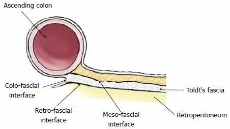
Figure 1 Depiction illustrating the relationships between the mesocolon, Toldt’s fascia (schematically exaggerated for descriptive purpose) and the retroperitoneum.
Meso-fascial interface is the apposition between the Toldt’s fascia and the overlaying mesocolon; Retro-fascial interface is the apposition between the Toldt’s fascia and the underlying retroperitoneum.
Figure 2 Depiction illustrating the meso-fascial (A) and retro-fascial (B) separation performed in the medial to lateral approach for laparoscopic right meso-colectomy.
In Meso-fascial separation (A), the plane of dissection lies between the inferior leaf of the mesocolon and the underlying Toldt’s fascia (schematically exaggerated for descriptive purpose), separating both components of the meso-fascial interface. In Retro-fascial separation (B), the plane of dissection lies between the Toldt’s fascia and the posterior parietal peritoneum covering the retroperitoneum, separating both components of the retro-fascial interface. Both dissections end with colo-fascial separation.
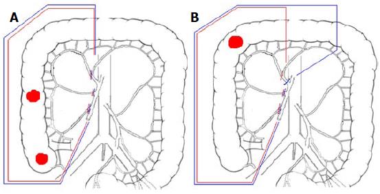
Figure 3 Schematic drawing illustrating the difference between extent of colon resection and lymph node harvesting between D3 right hemicolectomy accordingly to 2010 JSCCR guidelines (red lines) and complete mesocolic excision with central vascular ligation accordingly to Hohenberger’s rules (blue lines).
A: Cancer located in the caecum or ascending colon; B: Cancer located in the right (hepatic) flexure.
- Citation: Siani LM, Garulli G. Laparoscopic complete mesocolic excision with central vascular ligation in right colon cancer: A comprehensive review. World J Gastrointest Surg 2016; 8(2): 106-114
- URL: https://www.wjgnet.com/1948-9366/full/v8/i2/106.htm
- DOI: https://dx.doi.org/10.4240/wjgs.v8.i2.106


