Copyright
©The Author(s) 2016.
World J Gastrointest Surg. Dec 27, 2016; 8(12): 770-778
Published online Dec 27, 2016. doi: 10.4240/wjgs.v8.i12.770
Published online Dec 27, 2016. doi: 10.4240/wjgs.v8.i12.770
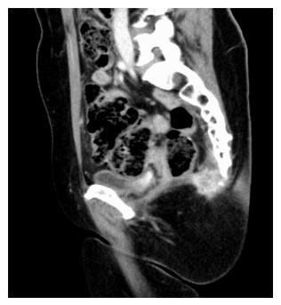
Figure 1 Radiological aspect of a local relapse infiltrating the coccix and lower sacral bone.
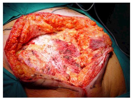
Figure 2 Preparation of skin flaps allows a complete exposure of maximus gluteus muscles.
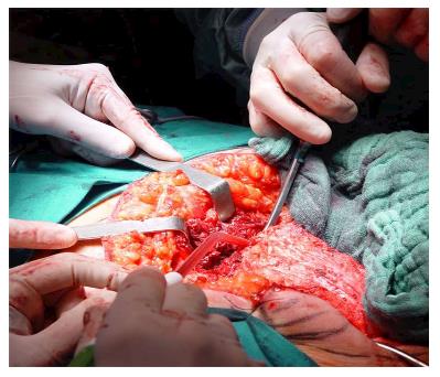
Figure 3 After the level of sacral transaction is identified the sacrum is osteotomized using normally a proper hammer and scalpel.
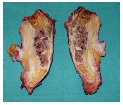
Figure 4 The figure shows a section of sacral specimen after S2 osteotomy.
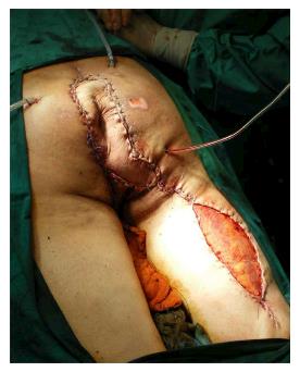
Figure 5 Example of a complex plastic reconstruction of the sacral area by a pedicled musculocutaneous flap and a thigh thin graft.
- Citation: Belli F, Gronchi A, Corbellini C, Milione M, Leo E. Abdominosacral resection for locally recurring rectal cancer. World J Gastrointest Surg 2016; 8(12): 770-778
- URL: https://www.wjgnet.com/1948-9366/full/v8/i12/770.htm
- DOI: https://dx.doi.org/10.4240/wjgs.v8.i12.770


