Copyright
©The Author(s) 2015.
World J Gastrointest Surg. Jul 27, 2015; 7(7): 123-127
Published online Jul 27, 2015. doi: 10.4240/wjgs.v7.i7.123
Published online Jul 27, 2015. doi: 10.4240/wjgs.v7.i7.123
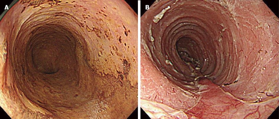
Figure 1 Endoscopic findings in case 1.
A: Before endoscopic submucosal dissection (ESD). The lesion covered three-quarters of the circumference; B: After ESD. We injected a steroid into an artificial ulcer.
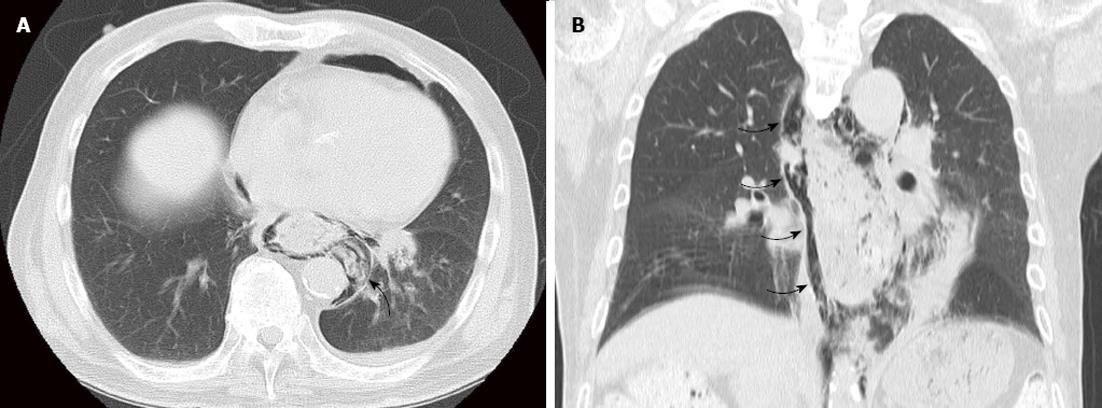
Figure 2 Enhanced computed tomography showed food residue in his mediastinum (arrow) (A) and mediastinal emphysema (arrow) (B).
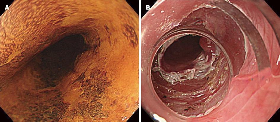
Figure 3 Endoscopic findings in case 2.
A: Before endoscopic submucosal dissection (ESD). The extent of the lesion ranged over half of its circumference; B: After ESD. We could not completely resect the lesion due to marked fibrosis.
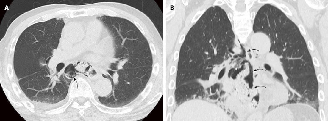
Figure 4 Enhanced computed tomography showed food residue in his mediastinum (arrow) (A) and mediastinal emphysema (arrow) (B).
- Citation: Matsuda Y, Kataoka N, Yamaguchi T, Tomita M, Sakamoto K, Makimoto S. Delayed esophageal perforation occurring with endoscopic submucosal dissection: A report of two cases. World J Gastrointest Surg 2015; 7(7): 123-127
- URL: https://www.wjgnet.com/1948-9366/full/v7/i7/123.htm
- DOI: https://dx.doi.org/10.4240/wjgs.v7.i7.123


