Copyright
©The Author(s) 2015.
World J Gastrointest Surg. Jun 27, 2015; 7(6): 98-101
Published online Jun 27, 2015. doi: 10.4240/wjgs.v7.i6.98
Published online Jun 27, 2015. doi: 10.4240/wjgs.v7.i6.98
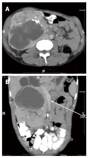
Figure 1 Computed tomography abdomen showing tumour encasing second part of duodenum and dilated common bile duct.
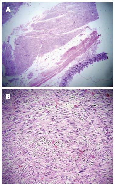
Figure 2 Microscopic findings (hematoxilin-eosin).
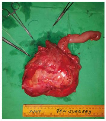
Figure 3 Gross specimen showing tumour.
- Citation: Bhambare MR, Pandya JS, Waghmare SB, Shetty TS. Gastrointestinal stromal tumour presenting as palpable abdominal mass: A rare entity. World J Gastrointest Surg 2015; 7(6): 98-101
- URL: https://www.wjgnet.com/1948-9366/full/v7/i6/98.htm
- DOI: https://dx.doi.org/10.4240/wjgs.v7.i6.98


