Copyright
©The Author(s) 2015.
World J Gastrointest Surg. May 27, 2015; 7(5): 82-85
Published online May 27, 2015. doi: 10.4240/wjgs.v7.i5.82
Published online May 27, 2015. doi: 10.4240/wjgs.v7.i5.82
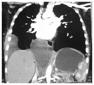
Figure 1 Computed tomography thorax and abdomen showing well defined thoraco-abdominal cyst.
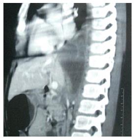
Figure 2 Lateral view of computed tomography thorax and abdomen showing extension of cyst in posterior mediastinum and abdomen.
Figure 3 Barium study-showing smooth indentation li lower esophagus with right sided.
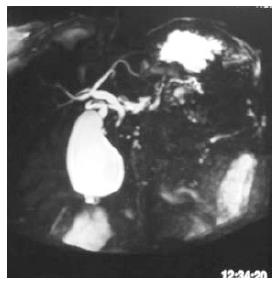
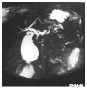
Figure 4 Magnetic resonance cholangiopancreatography, showing mild dilatation of multiple side branches of pancreatic duct in tail region without any peripancreatic collection.
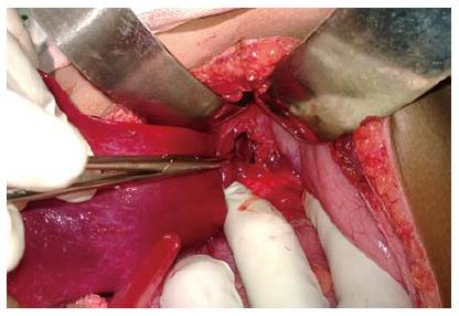
Figure 5 Intra operative photograph showing opening made in the anterior wall of pseudocyst.
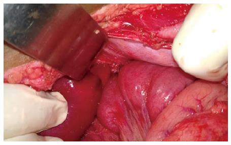
Figure 6 Intraoperative photograph showing completed anastomosis, Roux-en-Y loop seen behind stomach.
- Citation: Kamble RS, Gupta R, Gupta AR, Kothari PR, Dikshit KV, Kekre GA, Patil PS. Thoracoabdominal pseudocyst of pancreas: An rare location, managed by retrocolic retrogastric Roux-en-Y cystojejunostomy. World J Gastrointest Surg 2015; 7(5): 82-85
- URL: https://www.wjgnet.com/1948-9366/full/v7/i5/82.htm
- DOI: https://dx.doi.org/10.4240/wjgs.v7.i5.82


