Copyright
©The Author(s) 2015.
World J Gastrointest Surg. Apr 27, 2015; 7(4): 52-59
Published online Apr 27, 2015. doi: 10.4240/wjgs.v7.i4.52
Published online Apr 27, 2015. doi: 10.4240/wjgs.v7.i4.52
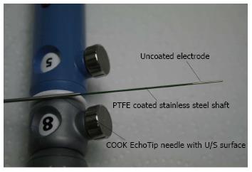
Figure 1 Close up of the HabibTM endoscopic ultrasound-radiofrequency ablation catheter showing uncoated electrode at the tip and the PTFE Coated stainless steel shaft.
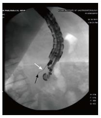
Figure 2 Fluoroscopic view of HabibTM endoscopic ultrasound-radiofrequency ablation catheter (black arrow) protruding out of the endoscopic ultrasound Biopsy needle (white arrow).
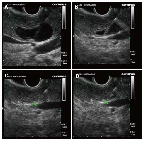
Figure 3 Endoscopic ultrasound pictures of radiofrequency ablation of pancreatic cyst.
A: Pancreatic cyst with the biopsy needle in position; B: Aspiration of the pancreatic cyst; C and D: Complete aspiration of the cyst followed by radiofrequency ablation using the endoscopic ultrasound radiofrequency ablation catheter.
Figure 4 Endoscopic ultrasound Pictures of radiofrequency ablation of pancreatic cyst.
A: Pancreatic cyst Pre ablation (arrow); B: Pancreatic cyst aspirated completed and the radiofrequency ablation with in process using the endoscopic ultrasound radiofrequency ablation catheter (arrow).
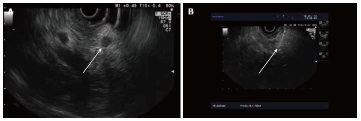
Figure 5 Endoscopic ultrasound radiofrequency ablation of pancreatic neuroendocrine tumors.
A and B: Endoscopic ultrasound pictures of the pancreatic neuroendocrine tumors pre and during ablation.
- Citation: Pai M, Habib N, Senturk H, Lakhtakia S, Reddy N, Cicinnati VR, Kaba I, Beckebaum S, Drymousis P, Kahaleh M, Brugge W. Endoscopic ultrasound guided radiofrequency ablation, for pancreatic cystic neoplasms and neuroendocrine tumors. World J Gastrointest Surg 2015; 7(4): 52-59
- URL: https://www.wjgnet.com/1948-9366/full/v7/i4/52.htm
- DOI: https://dx.doi.org/10.4240/wjgs.v7.i4.52


