Copyright
©The Author(s) 2015.
World J Gastrointest Surg. Mar 27, 2015; 7(3): 25-32
Published online Mar 27, 2015. doi: 10.4240/wjgs.v7.i3.25
Published online Mar 27, 2015. doi: 10.4240/wjgs.v7.i3.25
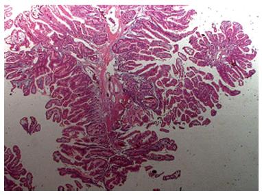
Figure 1 Histological features of a Peutz-Jeghers polyp.
Note that they are typically multilobulated with a papillary surface and branching bands of smooth muscle covered by hyperplastic glandular mucosa.
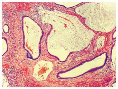
Figure 2 A Juvenile Polyp exhibiting a normal epithelium with a dense stroma, an inflammatory infiltrate and a smooth surface with dilated, mucus-filled cystic glands in the lamina propria.
Figure 3 Mucocutaneous pigmentation in Peutz-Jeghers Syndrome.
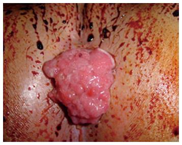
Figure 4 Prolapsed polyp through the anus in a patient with Juvenile Polyposis.
Figure 5 Feet queratosis (A), multiple facial triquilemomas (B) and oral mucosa papilomatosis (C) in a patients with Cowden’s Syndrome.
- Citation: Campos FG, Figueiredo MN, Martinez CAR. Colorectal cancer risk in hamartomatous polyposis syndromes. World J Gastrointest Surg 2015; 7(3): 25-32
- URL: https://www.wjgnet.com/1948-9366/full/v7/i3/25.htm
- DOI: https://dx.doi.org/10.4240/wjgs.v7.i3.25


