Copyright
©The Author(s) 2015.
World J Gastrointest Surg. Feb 27, 2015; 7(2): 21-24
Published online Feb 27, 2015. doi: 10.4240/wjgs.v7.i2.21
Published online Feb 27, 2015. doi: 10.4240/wjgs.v7.i2.21
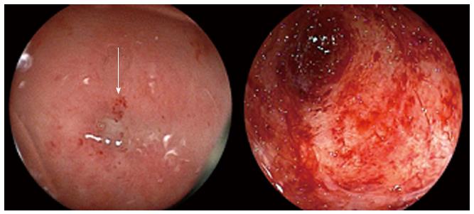
Figure 1 Colonoscopy through the ileostomy showed a tight stricture of the sigmoid colon at the anastomotic site (arrow).
The mucosa of the sigmoid colon was severely atrophic (right panel).
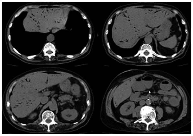
Figure 2 Computed tomography scan of the abdomen showed a marked amount of air throughout the portal venous system.
The transverse colon was dilated (arrow).
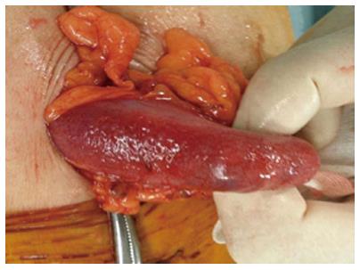
Figure 3 Intraoperative findings.
The transverse colon was edematous.
- Citation: Sadatomo A, Koinuma K, Kanamaru R, Miyakura Y, Horie H, Lefor AT, Yasuda Y. Hepatic portal venous gas after endoscopy in a patient with anastomotic obstruction. World J Gastrointest Surg 2015; 7(2): 21-24
- URL: https://www.wjgnet.com/1948-9366/full/v7/i2/21.htm
- DOI: https://dx.doi.org/10.4240/wjgs.v7.i2.21


