Copyright
©2014 Baishideng Publishing Group Inc.
World J Gastrointest Surg. Jul 27, 2014; 6(7): 142-145
Published online Jul 27, 2014. doi: 10.4240/wjgs.v6.i7.142
Published online Jul 27, 2014. doi: 10.4240/wjgs.v6.i7.142
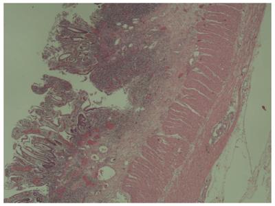
Figure 1 Biopsy from colonoscopy which revealed mucosal inflammation, compatible with ulcerative colitis.
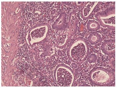
Figure 2 Histopathological image from the excised colon, typical of ulcerative colitis.
This image demonstrates marked lymphocytic infiltration (blue/purple) of the intestinal mucosa and architectural distortion of the crypts (right side of the image). The inflammation is shallow and affects only the mucosa sparing the muscularis mucosal (left side).
- Citation: Papaconstantinou I, Stefanopoulos A, Papailia A, Zeglinas C, Georgopoulos I, Michopoulos S. Isotretinoin and ulcerative colitis: A case report and review of the literature. World J Gastrointest Surg 2014; 6(7): 142-145
- URL: https://www.wjgnet.com/1948-9366/full/v6/i7/142.htm
- DOI: https://dx.doi.org/10.4240/wjgs.v6.i7.142


