Copyright
©2014 Baishideng Publishing Group Inc.
World J Gastrointest Surg. Jul 27, 2014; 6(7): 136-141
Published online Jul 27, 2014. doi: 10.4240/wjgs.v6.i7.136
Published online Jul 27, 2014. doi: 10.4240/wjgs.v6.i7.136
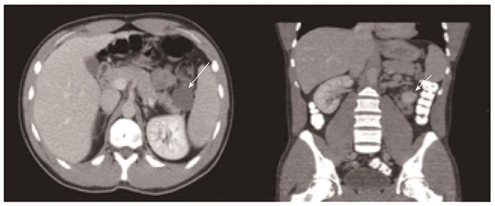
Figure 1 The usual appearance of lymphoepithelial cysts and lymphangiomas on contrast enhanced computerized tomography is that of simple cysts (arrows).
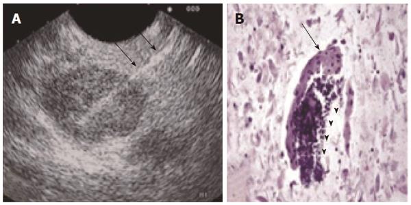
Figure 2 Endoscopic ultrasound guided fine needle aspiration (A) of the cyst with a 22G needle (black arrows) can aid in the correct diagnosis by demonstrating cellular elements characteristic of a lymphoepithelial cyst of the pancreas (B), including squamous (black arrow) and lymphoid subsets (black arrowheads) (HE stain, cell block preparation).
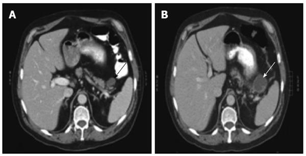
Figure 3 Lymphoepithelial cyst of the pancreas may increase in size during observation.
Contrast enhanced computed tomography images demonstrate that the cyst increased from 2.6 cm (A, black arrow) to 4.2 cm (B, white arrow) over a two-month period.
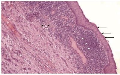
Figure 4 Lymphoepithelial cyst of the pancreas lined by squamous epithelium (black arrows), underlying lymphoid tissue (white arrowheads).
Pancreatic ductules are also identified (black arrowheads) (HE stain).
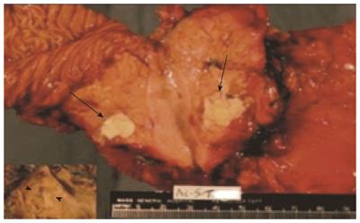
Figure 5 Photo of a transected lymphoepithelial cyst.
The characteristic keratinaceous cheesy material is shown (arrows). Inset: the multilocular cyst (arrowheads).
- Citation: Konstantinidis IT, Kambadakone A, Catalano OA, Sahani DV, Deshpande V, Forcione DG, Wargo JA, Fernandez-del Castillo C, Lillemoe KD, Warshaw AL, Ferrone CR. Lymphoepithelial cysts and cystic lymphangiomas: Under-recognized benign cystic lesions of the pancreas. World J Gastrointest Surg 2014; 6(7): 136-141
- URL: https://www.wjgnet.com/1948-9366/full/v6/i7/136.htm
- DOI: https://dx.doi.org/10.4240/wjgs.v6.i7.136


