Copyright
©2014 Baishideng Publishing Group Inc.
World J Gastrointest Surg. Jun 27, 2014; 6(6): 112-116
Published online Jun 27, 2014. doi: 10.4240/wjgs.v6.i6.112
Published online Jun 27, 2014. doi: 10.4240/wjgs.v6.i6.112
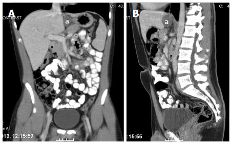
Figure 1 Computed tomography images of the esophageal duplication cyst (a).
A: Frontal view; B: Sagittal view.
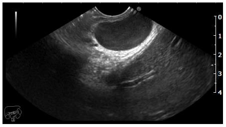
Figure 2 Endoscopic ultrasound image of the cyst.
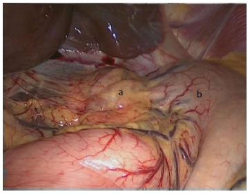
Figure 3 Preoperative image with the cyst (a) medial to the esophagus and stomach (b).
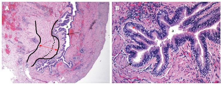
Figure 4 Histological findings of the cyst (hematoxylin/eosin staining).
A: Magnification × 20; B: Magnification × 40. Indicated are the lining of respiratory epithelium (black arrows) with a muscularis propia consisting of two muscular layers (red arrow), the lumen of the cyst (a) and the ciliated pseudo stratified columnar epithelium (red arrowhead).
- Citation: Castelijns PSS, Woensdregt K, Hoevenaars B, Nieuwenhuijzen GAP. Intra-abdominal esophageal duplication cyst: A case report and review of the literature. World J Gastrointest Surg 2014; 6(6): 112-116
- URL: https://www.wjgnet.com/1948-9366/full/v6/i6/112.htm
- DOI: https://dx.doi.org/10.4240/wjgs.v6.i6.112


