Copyright
©2014 Baishideng Publishing Group Co.
World J Gastrointest Surg. Apr 27, 2014; 6(4): 77-79
Published online Apr 27, 2014. doi: 10.4240/wjgs.v6.i4.77
Published online Apr 27, 2014. doi: 10.4240/wjgs.v6.i4.77
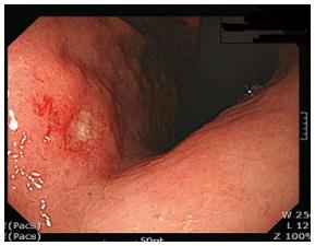
Figure 1 Endoscopic findings.
Ulcerative lesion in the lesser curvature of the lower body.
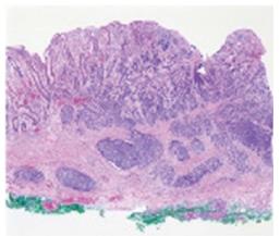
Figure 2 Endoscopic submucosal dissection specimen.
A hypercellular lesion was detected in the mucosa and submucosal layers.
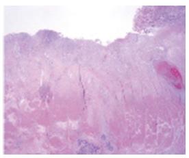
Figure 3 Laparoscopic assisted distal gastrectomy specimen.
The ulcerative lesion due to mucosal detachment after endoscopic submucosal dissection is distinguished from normal mucosa (right side). Fibrosis was observed in the submucosal layer and a hypercellular lesion that was the same as the endoscopic submucosal dissection specimen in the muscle and subserosa layers.
- Citation: Kang SH, Kim KH, Seo SH, An MS, Ha TK, Park HK, Bae KB, Choi CS, Oh SH, Choi YK. Neuroendocrine carcinoma of the stomach: A case report. World J Gastrointest Surg 2014; 6(4): 77-79
- URL: https://www.wjgnet.com/1948-9366/full/v6/i4/77.htm
- DOI: https://dx.doi.org/10.4240/wjgs.v6.i4.77


