Copyright
©2014 Baishideng Publishing Group Co.
World J Gastrointest Surg. Apr 27, 2014; 6(4): 74-76
Published online Apr 27, 2014. doi: 10.4240/wjgs.v6.i4.74
Published online Apr 27, 2014. doi: 10.4240/wjgs.v6.i4.74
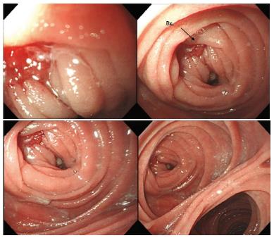
Figure 1 Endoscopic findings.
There was a medium-sized single small polypoid infiltrative ill-defined mass, with nodular overlying mucosa without bleeding evidence at jejunal pouch (1.2 cm in diameter). Tubular adenocarcinoma, well differentiated.
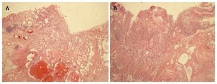
Figure 2 Pathological findings.
A: January 2008, slide of gastric cancer lesion (primary lesion); B: December 2011, slide of jejunal stump lesion (recurrent lesion).
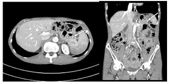
Figure 3 Pre-operation computed tomography findings.
No evidence of local tumor recurrence or distant metastasis. Arrow: Distal jejunal stump stapling line (recurrence site).
- Citation: Yoo JH, Seo SH, An MS, Ha TK, Kim KH, Bae KB, Choi CS, Oh SH, Choi YK. Recurrence of gastric cancer in the jejunal stump after radical total gastrectomy. World J Gastrointest Surg 2014; 6(4): 74-76
- URL: https://www.wjgnet.com/1948-9366/full/v6/i4/74.htm
- DOI: https://dx.doi.org/10.4240/wjgs.v6.i4.74


