Copyright
©2014 Baishideng Publishing Group Co.
World J Gastrointest Surg. Apr 27, 2014; 6(4): 70-73
Published online Apr 27, 2014. doi: 10.4240/wjgs.v6.i4.70
Published online Apr 27, 2014. doi: 10.4240/wjgs.v6.i4.70
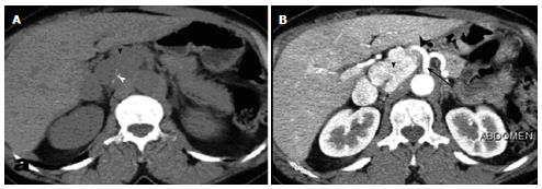
Figure 1 Abdominal computed tomography without (A) and after (B) contrast material administration, showing the tumor (arrowhead) with calcifications (white arrowhead) and precocious enhancement.
Note the close tumoral relationship to the celiac trunk (black arrow) and hepatic artery (black arrowhead).
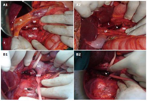
Figure 2 Operative view.
A: Patient 1. A1: Separation of the tumor (T) from the anterior aspect of the inferior vena cava (IVC); A2: Surgical site after tumor resection (arrowhead); B: Patient 3. B1: Separation of the tumor (T) from the posterior wall of the IVC; B2: Surgical site after tumor resection (arrowhead). LRV: Left renal vein; D: Duodenum; HP: Head of the pancreas; PL: Liver pedicle; RRV: Right renal vein; RK: Right kidney; HA: Hepatic artery; LP: Liver pedicle; L: Liver (Lobe of Spiegel).
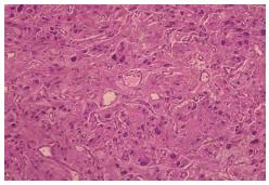
Figure 3 Microscopic view of paraganglioma.
Large polygonal cells with granular cytoplasm arranged in nests (hematoxylin and eosin).
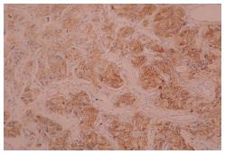
Figure 4 Immunohistochemistry.
Tumoral cells strongly express anti-chromogranin antibody.
- Citation: Kallel H, Hentati H, Baklouti A, Gassara A, Saadaoui A, Halek G, Landolsi S, Ouaer ME, Chaieb W, Maamouri F, Mannaï S. Retroperitoneal paragangliomas: Report of 4 cases. World J Gastrointest Surg 2014; 6(4): 70-73
- URL: https://www.wjgnet.com/1948-9366/full/v6/i4/70.htm
- DOI: https://dx.doi.org/10.4240/wjgs.v6.i4.70


