Copyright
©2014 Baishideng Publishing Group Co.
World J Gastrointest Surg. Apr 27, 2014; 6(4): 65-69
Published online Apr 27, 2014. doi: 10.4240/wjgs.v6.i4.65
Published online Apr 27, 2014. doi: 10.4240/wjgs.v6.i4.65
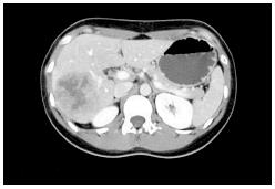
Figure 1 A computed tomography scan with nonionic contrast confirmed a mass located in the posterior right lobe within segments VI-VII and measuring 7.
2 cm × 6.0 cm. The lesion demonstrated peripheral enhancement with central necrosis but no evidence for portal vein invasion. The hepatic veins were patent and no biliary dilatation was observed. No pulmonary lesion was highlighted.
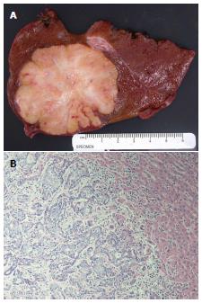
Figure 2 Photograph.
A: Right lobe liver resection specimen showing a 6.0 cm × 5.5 cm × 5 cm well-circumscribed tumor with a firm, heterogeneous, yellow tan and white cut surface with areas of fibrosis. The surrounding liver is unremarkable; B: Representative photomicrograph of the tumor shows anastomosing glandular structures composed of highly pleomorphic epithelial cells in a desmoplastic stroma. The findings are consistent with intrahepatic cholangiocarcinoma (HE, original magnification × 200).
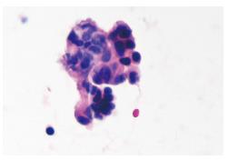
Figure 3 Endoscopic ultrasound-guided fine needle aspiration biopsy of pancreatic tail mass.
Cytospin material showing a loose three-dimensional cluster of cells with high nuclear to cytoplasmic ratio, hyperchromasia, and eosinophilic cytoplasm. Several other clusters similar to these were found on the slide. The findings are consistent with adenocarcinoma compatible with metastatic cholangiocarcinoma.
- Citation: Labgaa I, Carrasco-Avino G, Fiel MI, Schwartz ME. Pancreatic recurrence of intrahepatic cholangiocarcinoma: Case report and review of the literature. World J Gastrointest Surg 2014; 6(4): 65-69
- URL: https://www.wjgnet.com/1948-9366/full/v6/i4/65.htm
- DOI: https://dx.doi.org/10.4240/wjgs.v6.i4.65


