Copyright
©2014 Baishideng Publishing Group Co.
World J Gastrointest Surg. Mar 27, 2014; 6(3): 51-54
Published online Mar 27, 2014. doi: 10.4240/wjgs.v6.i3.51
Published online Mar 27, 2014. doi: 10.4240/wjgs.v6.i3.51
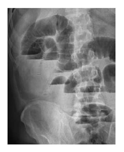
Figure 1 X-ray plain abdominal imaging (posteroanterior) revealed intestinal obstruction with marked small bowel air-fluid levels.
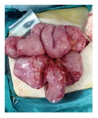
Figure 2 Intraoperative photographs taken along the midline incision and showing the encapsulated small bowel segments with a dense fibrous layer.
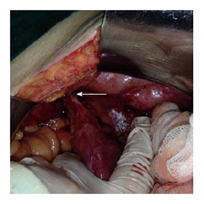
Figure 3 Intraoperative photographs showing the Meckel’s diverticulum incarcerated within the right inguinal canal (arrow).
- Citation: Akbulut S, Yagmur Y, Babur M. Coexistence of abdominal cocoon, intestinal perforation and incarcerated Meckel’s diverticulum in an inguinal hernia: A troublesome condition. World J Gastrointest Surg 2014; 6(3): 51-54
- URL: https://www.wjgnet.com/1948-9366/full/v6/i3/51.htm
- DOI: https://dx.doi.org/10.4240/wjgs.v6.i3.51


