Copyright
©2014 Baishideng Publishing Group Co.
World J Gastrointest Surg. Mar 27, 2014; 6(3): 42-46
Published online Mar 27, 2014. doi: 10.4240/wjgs.v6.i3.42
Published online Mar 27, 2014. doi: 10.4240/wjgs.v6.i3.42
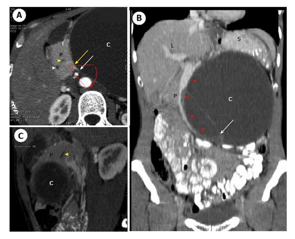
Figure 1 Preoperative contrast-enhanced computed tomography showing a huge cyst with septa developed close to the head of the pancreas, exophytic development to the left and downward, and rotation of the mesenteric axis.
A: Axial view showing the head of the P with the intrapancreatic main bile duct (white arrowhead) and ventral pancreatic duct (yellow arrowhead). There is rotation of the mesenteric axis, as shown by the oblique plane made by the superior mesenteric artery (white arrow) and the superior mesenteric vein (yellow arrow); B: Coronal view showing thin septa within the macrocyst (white arrow) and deviation without thrombosis of the mesenterico-portal axis (red arrowhead); C: Sagittal view showing anterior development of the cyst, close to the head of the pancreas (P) and the ventral pancreatic duct (yellow arrowhead). L: Liver; S: Spleen; P: Pancreas; C: Cyst.
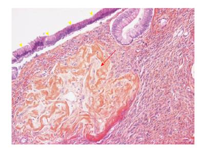
Figure 2 Histologic examination (× 20) showing the cyst lined by tall columnar epithelial cell (yellow arrowheads) with underlying ovarian-type stroma composed of densely packed spindle cells (red arrow).
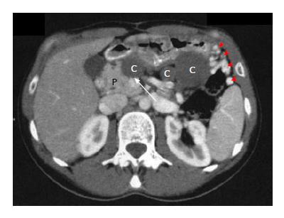
Figure 3 Post-operative contrast-enhanced computed tomography showing three low-density homogenous cystic lesions suggesting secondary dissemination of the resected cyst.
Major tributaries were also present around the stomach (red arrowheads). The P has a total distal atrophy as shown by lack of pancreatic tissue behind the mesenterico-portal axis (white arrow). P: Pancreas; C: Cyst.
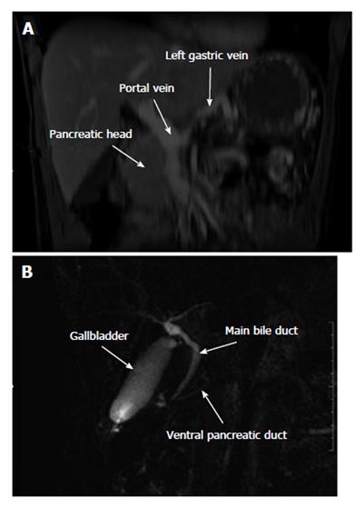
Figure 4 Coronal magnetic resonance imagery showing lack of the body and tail of the pancreas and of the splenic vein (A), magnetic resonance cholangiopancreatography showing the common bile duct joining the ventral pancreatic duct at the posterior part of the head of the pancreas (B).
The dorsal pancreatic duct is not visible.
- Citation: Gagnière J, Dupré A, Ines DD, Tixier L, Pezet D, Buc E. Giant mucinous cystic adenoma with pancreatic atrophy mimicking dorsal agenesis of the pancreas. World J Gastrointest Surg 2014; 6(3): 42-46
- URL: https://www.wjgnet.com/1948-9366/full/v6/i3/42.htm
- DOI: https://dx.doi.org/10.4240/wjgs.v6.i3.42


