Copyright
©2014 Baishideng Publishing Group Co.
World J Gastrointest Surg. Feb 27, 2014; 6(2): 33-37
Published online Feb 27, 2014. doi: 10.4240/wjgs.v6.i2.33
Published online Feb 27, 2014. doi: 10.4240/wjgs.v6.i2.33
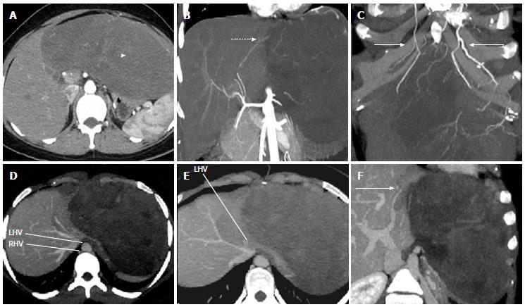
Figure 1 A 28-year-old lady with low grade fibromyxoid sarcoma on dynamic computed tomography.
A: Soft tissue homogenously enhancing mildly hypodense mass (arrowhead) in the region of left lobe of liver is seen on the arterial phase of the dynamic triple phase computed tomography scan; B: The mass shows small arterial feeders from the left hepatic artery (dotted arrow); C: Large feeders from bilateral internal mammary arteries (solid arrows) to the mass are seen; D: Middle hepatic vein and right hepatic vein show normal patency and course; E: Left hepatic vein is only seen at the ostium, appears compressed by the mass and is not seen beyond the ostium; F: Left portal vein (solid arrow) is also splayed and partially attenuated by the tumor.
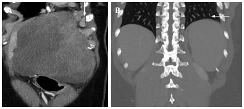
Figure 2 Sagittal and coronal multiplanar reformats of a 28-year-old lady with low grade fibromyxoid sarcoma on dynamic computed tomography.
A: Sagittal reconstruction of the abdomen shows supra-diaphragmatic enlarged lymph nodes (solid arrow); B: Coronal reconstruction images shows normal basal lung fields (dotted arrow).
Figure 3 Gross, histopathological and immunochemistry slides of 28-year-old lady with low grade fibromyxoid sarcoma.
A: Gross specimen of the resected tumor mass along with the left lower 3 ribs; B: Histopathology at (× 100, HE stain) showing low cellularity with bland appearing spindle and stellate cells and minimal nuclear atypia; C: Histopathology (× 100, HE) shows low grade fibromyxoid sarcoma with focal areas of storiform and fascicular arrangement of spindle cells; D: Spindle cells show strong positive immunostaining with vimentin (× 200, Vimentin, LSAB immunohistochemistry method).
- Citation: Thapar S, Ahuja A, Rastogi A. Rare diaphragmatic tumor mimicking liver mass. World J Gastrointest Surg 2014; 6(2): 33-37
- URL: https://www.wjgnet.com/1948-9366/full/v6/i2/33.htm
- DOI: https://dx.doi.org/10.4240/wjgs.v6.i2.33


