Copyright
©2014 Baishideng Publishing Group Inc.
World J Gastrointest Surg. Dec 27, 2014; 6(12): 248-252
Published online Dec 27, 2014. doi: 10.4240/wjgs.v6.i12.248
Published online Dec 27, 2014. doi: 10.4240/wjgs.v6.i12.248
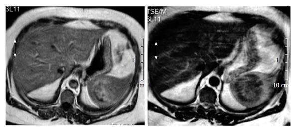
Figure 1 Magnetic resonance imaging shows a mass of 6 cm in diameter.
Axial T1W image shows well circumscribed solid and heterogeneous intrasplenic mass. It might seem to have an excentric scar although calcification could also be possible. It is difficult to discern by magnetic resonance imaging.
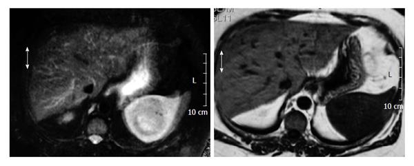
Figure 2 Axial T2W fat sat image shows a large intrasplenic mass.
Notice the slightly decrease of signal intensity and the lack of a peripherical capsule.
Figure 3 Histopathological study.
A: Section of the spleen with a inflammatory pseudotumor (20 × HE); B: High power xamination revealed a fibroblastic and myofibroblastic proliferation with lymphocytes and plasma cells (40 × HE); C: Several tuberculoid granuloma were identified in the spleen and and in the hilar lymph node (20 × HE).
- Citation: Prieto-Nieto MI, Pérez-Robledo JP, Díaz-San Andrés B, Nistal M, Rodríguez-Montes JA. Inflammatory pseudotumour of the spleen associated with splenic tuberculosis. World J Gastrointest Surg 2014; 6(12): 248-252
- URL: https://www.wjgnet.com/1948-9366/full/v6/i12/248.htm
- DOI: https://dx.doi.org/10.4240/wjgs.v6.i12.248


