Copyright
©2014 Baishideng Publishing Group Inc.
World J Gastrointest Surg. Oct 27, 2014; 6(10): 204-207
Published online Oct 27, 2014. doi: 10.4240/wjgs.v6.i10.204
Published online Oct 27, 2014. doi: 10.4240/wjgs.v6.i10.204
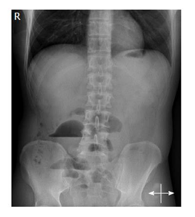
Figure 1 X-ray shows air-fluid levels in projection of the small bowel.
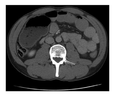
Figure 2 Computed tomography-scan shows distended small bowel loops and in the right mid-abdomen 11 cm × 6.
4 cm × 7.8 cm fluid and gas containing cavity with thickened wall.
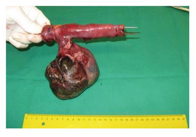
Figure 3 Torsioned necrotic Meckel´s diverticle obstructing the adjacent small bowel.
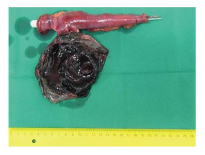
Figure 4 The mucosa of the diverticle was entirely necrotic, the diverticle was filled with hemorrhagic fluid.
- Citation: Murruste M, Rajaste G, Kase K. Torsion of Meckel's diverticulum as a cause of small bowel obstruction: A case report. World J Gastrointest Surg 2014; 6(10): 204-207
- URL: https://www.wjgnet.com/1948-9366/full/v6/i10/204.htm
- DOI: https://dx.doi.org/10.4240/wjgs.v6.i10.204


