Copyright
©2013 Baishideng Publishing Group Co.
World J Gastrointest Surg. Jun 27, 2013; 5(6): 199-201
Published online Jun 27, 2013. doi: 10.4240/wjgs.v5.i6.199
Published online Jun 27, 2013. doi: 10.4240/wjgs.v5.i6.199
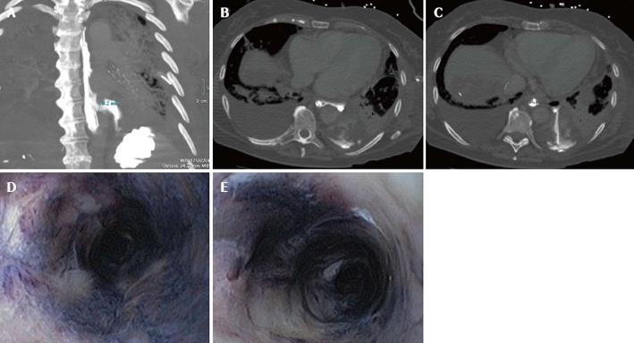
Figure 1 Computed tomography-thorax scan with contrast given via the nasogastric feeding tube.
A: A coronal coupe where the esophagus is seen to the left of the vertebral column and the perforation lights up two thirds of the esophagus with leakage of contrast into the pleural space; B and C: Transversal coupes of the thorax with contrast leakage via the esophagus into the pleural space; D and E: A circumferentially black-appearing esophagus at endoscopy.
- Citation: Groenveld RL, Bijlsma A, Steenvoorde P, Ozdemir A. A black perforated esophagus treated with surgery: Report of a case. World J Gastrointest Surg 2013; 5(6): 199-201
- URL: https://www.wjgnet.com/1948-9366/full/v5/i6/199.htm
- DOI: https://dx.doi.org/10.4240/wjgs.v5.i6.199


