Copyright
©2013 Baishideng Publishing Group Co.
World J Gastrointest Surg. Jun 27, 2013; 5(6): 192-194
Published online Jun 27, 2013. doi: 10.4240/wjgs.v5.i6.192
Published online Jun 27, 2013. doi: 10.4240/wjgs.v5.i6.192
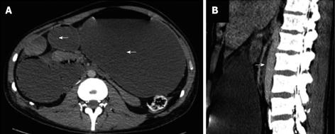
Figure 1 Contrast computed tomography revealing.
A: A complete gastric outlet obstruction (white arrow); B: With distension of the stomach, and the duodenum, with a flat bowel after its pass behind the superior mesenteric artery (white arrow).
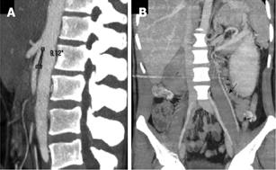
Figure 2 Contrast computed tomography with vascular reconstruction revealing.
A: That the aortomesenteric angle was decreased to 9; B: A left renal vein obstruction with left gonadal vein dilatation.
- Citation: Jeune F, d’Assignies G, Sauvanet A, Gaujoux S. A rare cause of obstructive jaundice and gastric outlet obstruction. World J Gastrointest Surg 2013; 5(6): 192-194
- URL: https://www.wjgnet.com/1948-9366/full/v5/i6/192.htm
- DOI: https://dx.doi.org/10.4240/wjgs.v5.i6.192


