Copyright
©2013 Baishideng Publishing Group Co.
World Journal of Gastrointestinal Surgery. Nov 27, 2013; 5(11): 306-308
Published online Nov 27, 2013. doi: 10.4240/wjgs.v5.i11.306
Published online Nov 27, 2013. doi: 10.4240/wjgs.v5.i11.306
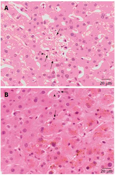
Figure 1 Active Epstein-Barr virus infection with hemophagocytosis not with lymphoma.
A: Large histiocytes showing erythrophagocytosis (arrows) and leucophagocytosis (black arrowhead); B: A Kupffer cell (white arrowhead) and a large histiocyte (arrowhead) have phagocytosed a lymphocyte (arrow), moderate cholestasis is present (white arrow).
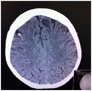
Figure 2 Focal lesion anteriorly in the left parietal lobe.
- Citation: Virdis F, Tacci S, Messina F, Varcada M. Hemophagocytic lymphohistiocytosis caused by primary Epstein-Barr virus in patient with Crohn’s disease. World Journal of Gastrointestinal Surgery 2013; 5(11): 306-308
- URL: https://www.wjgnet.com/1948-9366/full/v5/i11/306.htm
- DOI: https://dx.doi.org/10.4240/wjgs.v5.i11.306


