Copyright
©2013 Baishideng Publishing Group Co.
World J Gastrointest Surg. Oct 27, 2013; 5(10): 272-277
Published online Oct 27, 2013. doi: 10.4240/wjgs.v5.i10.272
Published online Oct 27, 2013. doi: 10.4240/wjgs.v5.i10.272
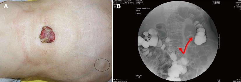
Figure 1 Findings of diagnostic nodule and colonorectal enema.
A: A 5.0- × 4.5-cm hard, ulcerated, caulescent tumor was observed in the umbilicus. Two swollen right inguinal lymph nodes were palpable (circle); B: Colonorectal enema showed an apple core sign at the transverse colon near the hepatic flexure, red arrow: the length of narrowed lumen.
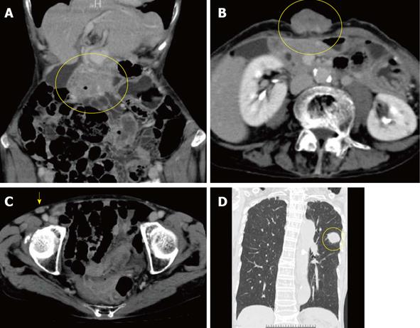
Figure 2 Computer tomography showed tumors of the transverse colon (A, circle), umbilicus (B, circle), right inguinal lymph nodes (C, arrow) and left lung (D, circle).
The lung tumor was accompanied by a typical spicula. Vessels along the round ligament of the liver did not develop to and from the umbilical tumor.
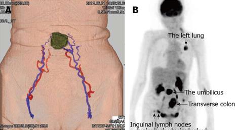
Figure 3 Findings of dynamic computer tomography and fluorodeoxyglucose-positron emission tomography.
A: Dynamic computer tomography revealed feeding arteries and drainage veins for the umbilical tumor were the inferior epigastric vessels; B: Fluorodeoxyglucose-positron emission tomography showed uptake in the transverse colon, umbilicus, right inguinal lymph nodes, and left lung (arrows).
Figure 4 Intraoperative findings and surgical procedures.
A: The umbilicus and caulescent tumor were resected en bloc with establishment of a surgical margin and ligation of vessels; B: Single-site laparoscopic surgery involving right hemicolectomy with paracolic lymph node dissection was subsequently performed. C: Indocyanine green was injected into the paraumbilical portion beforehand, allowing for detection of the drainage lymph nodes in the abdomen (arrow). SMNJ: Sister Mary Joseph’s nodule.
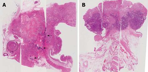
Figure 5 Histopathological findings of the primary colon cancer and Sister Mary Joseph’s nodule are shown (hematoxylin and eosin staining).
A: In the primary colon cancer, severe invasion and tumor thrombosis were observed in the vessels (circle), and nontortuous arteries and multiple A-V shunts were also present (arrows); B: The umbilical tumor was consistent with a moderately differentiated adenocarcinoma of colonic origin. The peritoneum of the umbilicus revealed normal findings.
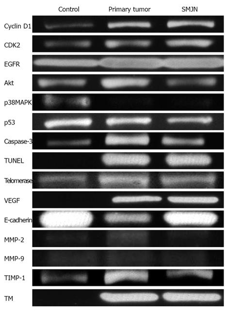
Figure 6 Actual intensities of the normal colon, primary colon tumor, and Sister Mary Joseph’s nodule in western blotting gelatin zymography, and real-time polymerase chain reaction (telomerase) are shown.
CDK2: Cyclin-dependent kinase 2; EGFR: Epidermal growth factor receptor; MAPK: Mitogen-activated protein kinase; TUNEL: Terminal deoxynucleotidyl transferase dUTP nick end labeling; VEGF: Vascular endothelial growth factor; MMP: Matrix metalloproteinase; TIMP: Tissue inhibitor of metalloproteinase; TM: Thrombomodulin; SMNJ: Sister Mary Joseph’s nodule.
- Citation: Hori T, Okada N, Nakauchi M, Hiramoto S, Kikuchi-Mizota A, Kyogoku M, Oike F, Sugimoto H, Tanaka J, Morikami Y, Shigemoto K, Ota T, Kaneko M, Nakatsuji M, Okae S, Tanaka T, Gunji D, Yoshioka A. Hematogenous umbilical metastasis from colon cancer treated by palliative single-incision laparoscopic surgery. World J Gastrointest Surg 2013; 5(10): 272-277
- URL: https://www.wjgnet.com/1948-9366/full/v5/i10/272.htm
- DOI: https://dx.doi.org/10.4240/wjgs.v5.i10.272


