Copyright
©2012 Baishideng Publishing Group Co.
World J Gastrointest Surg. Sep 27, 2012; 4(9): 220-222
Published online Sep 27, 2012. doi: 10.4240/wjgs.v4.i9.220
Published online Sep 27, 2012. doi: 10.4240/wjgs.v4.i9.220
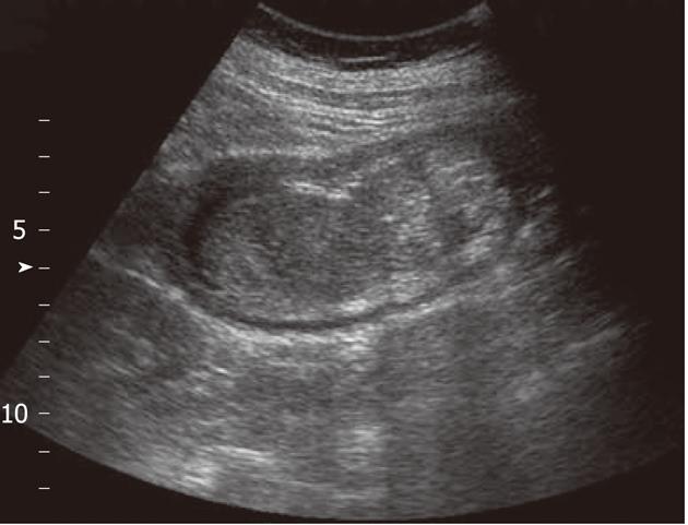
Figure 1 Abdominal echo showed prominent swelling of the intestinal wall with target signs and a hyperechoic mass of 67 mm × 60 mm in size over the right lower abdomen, and the impression was one of small-bowel intussusception with gangrenous change.
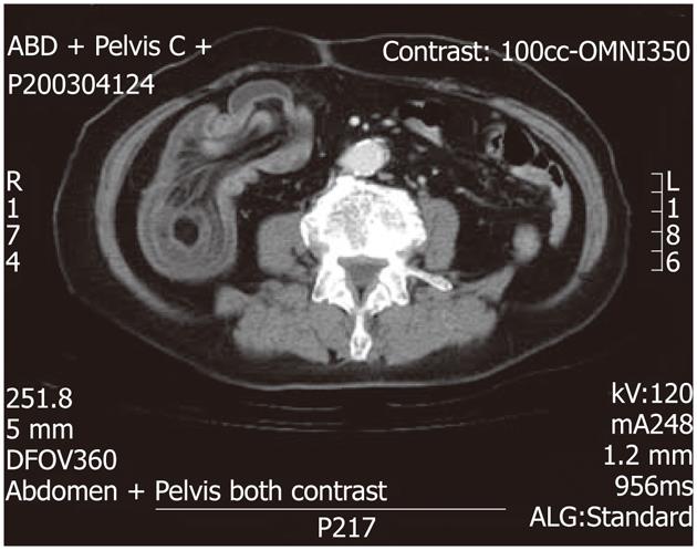
Figure 2 Computerized tomography showed ileocolic intussusception of the terminal ileum into the ascending colon with thickening of the bowel wall.
There are some fat-density lesions in the lumen of terminal ileum, highly suspected terminal ileum lipoma.
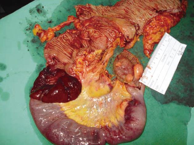
Figure 3 Macroscopic finding of excised specimen showed intussusception of terminal ileum caused by multiple submucosal nodules.
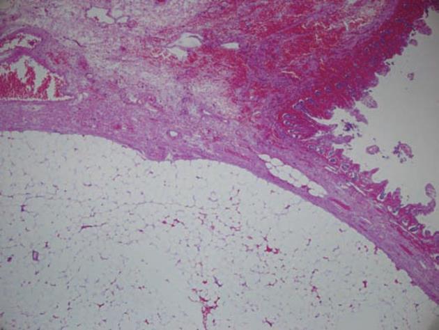
Figure 4 Microscopic finding of the ileal nodule showed submucosal lipoma composed of mature adipose tissue and surrounded by a fibrotic capsule.
Focal mucosal necrosis was also noted (HE, 40 ×).
- Citation: Hou YC, Lee PC, Chang JJ, Lai PS. Laparoscopic management of small-bowel intussusception in a 64-year-old female with ileoal lipomas. World J Gastrointest Surg 2012; 4(9): 220-222
- URL: https://www.wjgnet.com/1948-9366/full/v4/i9/220.htm
- DOI: https://dx.doi.org/10.4240/wjgs.v4.i9.220


