Copyright
©2012 Baishideng.
World J Gastrointest Surg. Jul 27, 2012; 4(7): 185-189
Published online Jul 27, 2012. doi: 10.4240/wjgs.v4.i7.185
Published online Jul 27, 2012. doi: 10.4240/wjgs.v4.i7.185
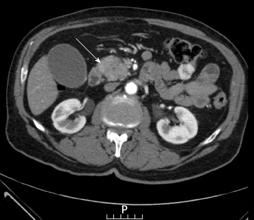
Figure 1 A computed tomography scan sagittal slide showing thickening of the pancreatic head with hypo-dense center (arrow) and with no obvious acinari structure.
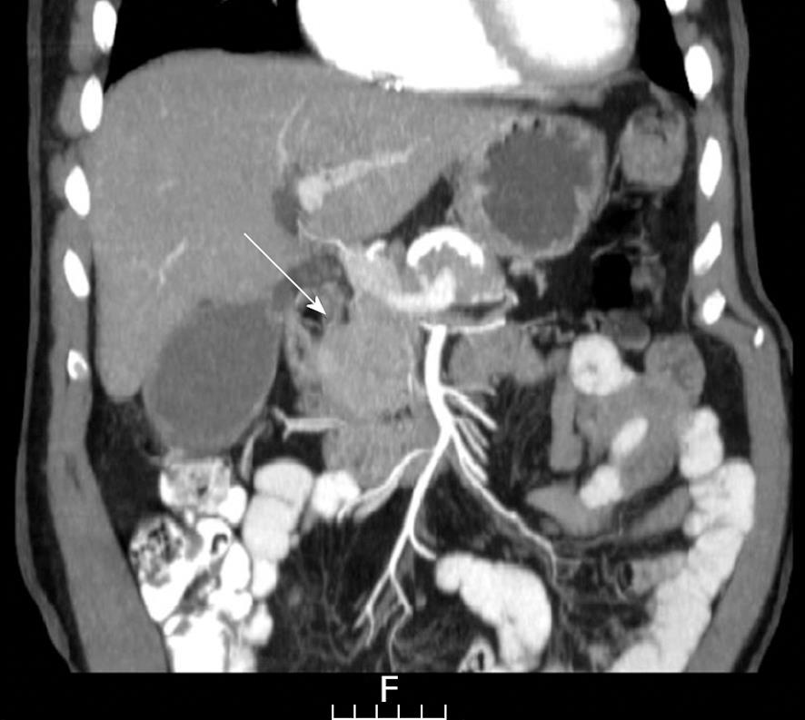
Figure 2 A coronal reconstruction reinforcing the sagittal finding (arrow).
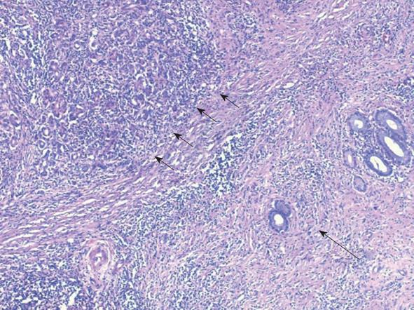
Figure 3 Chronic inflammation with diffuse fibrosis of the acini (short arrows) and around the small pancreatic ducts (long arrow); HE, × 50.
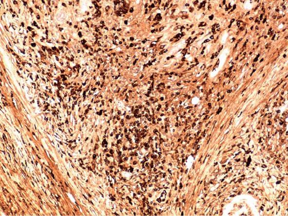
Figure 4 Numerous plasma cells, positive for IgG4 in inflammatory infiltrate; immunoperoxidase, × 100.
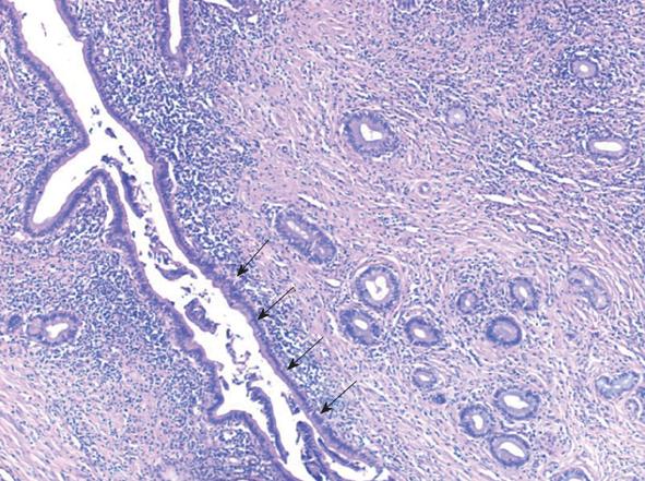
Figure 5 Marked fibrosis and lymphoplasmacytoid infiltration around the middle sized pancreatic ducts showing low grade pancreatic intraepithelial neoplasia (arrows); HE, × 50.
- Citation: Brauner E, Lachter J, Ben-Ishay O, Vlodavsky E, Kluger Y. Autoimmune pancreatitis misdiagnosed as a tumor of the head of the pancreas. World J Gastrointest Surg 2012; 4(7): 185-189
- URL: https://www.wjgnet.com/1948-9366/full/v4/i7/185.htm
- DOI: https://dx.doi.org/10.4240/wjgs.v4.i7.185


