Copyright
©2012 Baishideng.
World J Gastrointest Surg. Jun 27, 2012; 4(6): 157-162
Published online Jun 27, 2012. doi: 10.4240/wjgs.v4.i6.157
Published online Jun 27, 2012. doi: 10.4240/wjgs.v4.i6.157
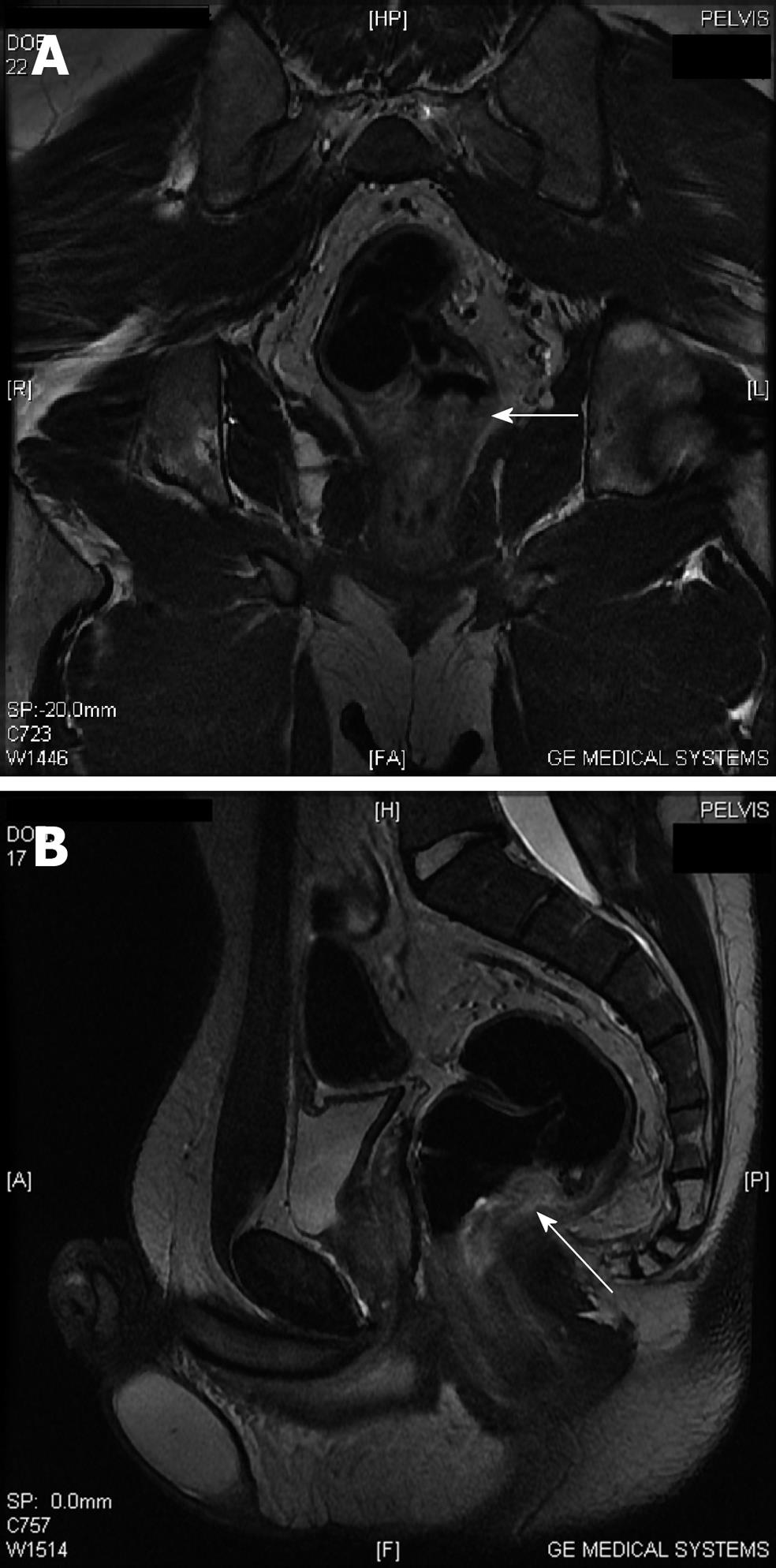
Figure 1 Coronal (A) and sagittal (B) T2 sequence MRI demonstrating the polyp (arrows).
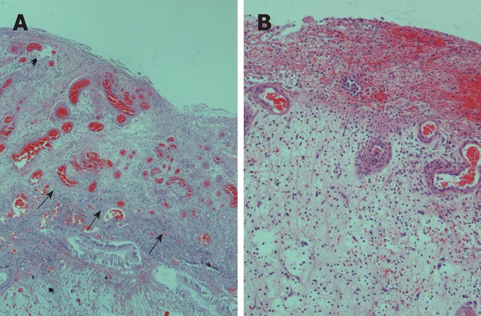
Figure 2 A cap of inflamed and ulcerated granulation tissue was observed in most superficial regions (arrows) (A), covered by fibrin and inflammatory exudate, as presented in higher magnification in (B).
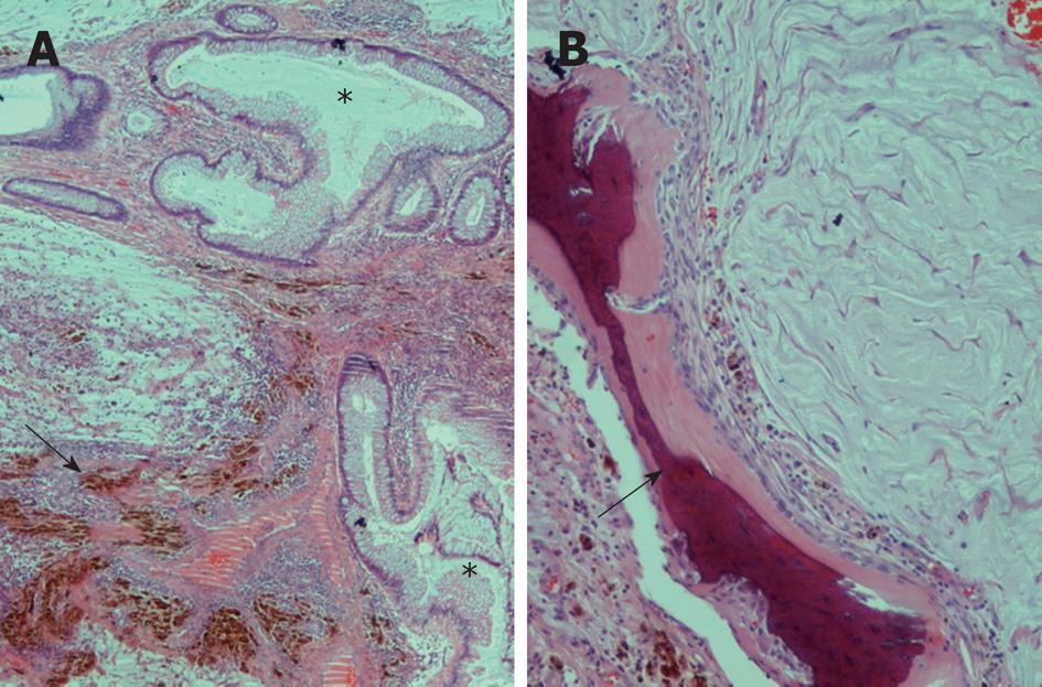
Figure 3 Dilated or tortuous colonic crypts (asterisks) were observed in central parts, alternating with lakes of mucin and hemosiderin deposits (arrow) in fibrotic or inflammatory areas (A), and heterotopic bone formation (arrow) was observed focally, adjacent to mucin lakes (B).
- Citation: Papaconstantinou I, Karakatsanis A, Benia X, Polymeneas G, Kostopoulou E. Solitary rectal cap polyp: Case report and review of the literature. World J Gastrointest Surg 2012; 4(6): 157-162
- URL: https://www.wjgnet.com/1948-9366/full/v4/i6/157.htm
- DOI: https://dx.doi.org/10.4240/wjgs.v4.i6.157


