Copyright
©2012 Baishideng.
World J Gastrointest Surg. May 27, 2012; 4(5): 104-113
Published online May 27, 2012. doi: 10.4240/wjgs.v4.i5.104
Published online May 27, 2012. doi: 10.4240/wjgs.v4.i5.104
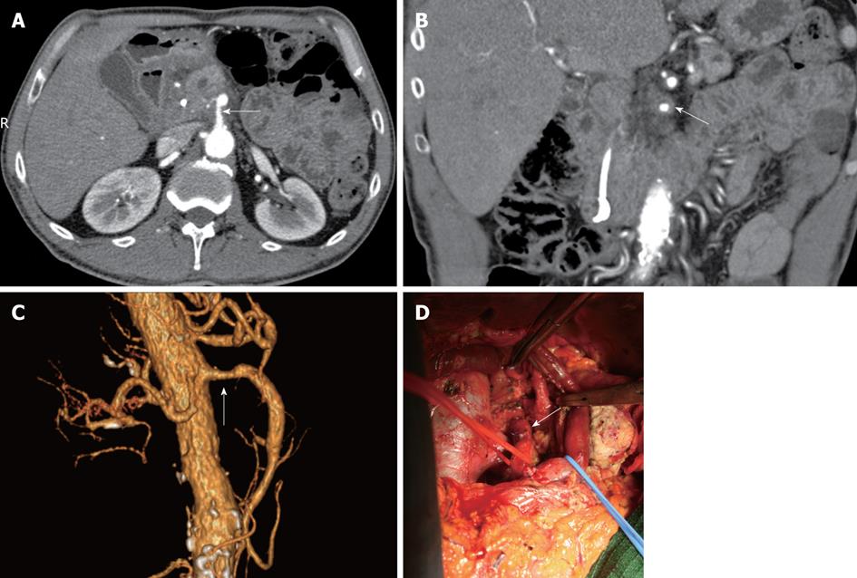
Figure 1 Pancreatic head carcinoma.
A: Axial image showed that the superior mesenteric artery (SMA) was surrounded by more than 50% of the vessel (white arrow) circumference by tumor and the vessel wall appeared infiltrated; B: Coronal oblique plane. Computed tomography image showed tumor encasement (white arrow) of the SMA (more than 180° of the vessel circumference surrounded by tumor); C: Volume rendering (VR) 3D images showed and a segment of the SMA stenosed (white arrow); D: Extended pancreaticoduodenectomy (Whipple procedure) with the pancreatic body excision and superior mesenteric vein resection. At surgical exploration, the common hepatic artery (white arrow) was found not to be invaded by tumor and tumor was successfully resected.
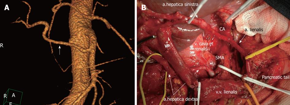
Figure 2 Michel’s type II celiac-mesenterial anatomy.
A: 3D computed tomography angiographic image. Replaced right hepatic artery (white arrow); B: View of the operating field after the extended pancreaticoduodenectomy. Note a the presence of a replaced right hepatic artery originating from the superior mesenteric artery (SMA). CA: Celiac artery.
- Citation: Zakharova OP, Karmazanovsky GG, Egorov VI. Pancreatic adenocarcinoma: Outstanding problems. World J Gastrointest Surg 2012; 4(5): 104-113
- URL: https://www.wjgnet.com/1948-9366/full/v4/i5/104.htm
- DOI: https://dx.doi.org/10.4240/wjgs.v4.i5.104


