Copyright
©2012 Baishideng Publishing Group Co.
World J Gastrointest Surg. Feb 27, 2012; 4(2): 41-44
Published online Feb 27, 2012. doi: 10.4240/wjgs.v4.i2.41
Published online Feb 27, 2012. doi: 10.4240/wjgs.v4.i2.41
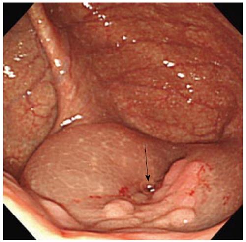
Figure 1 Lesion was beside the appendix orifice (arrow).
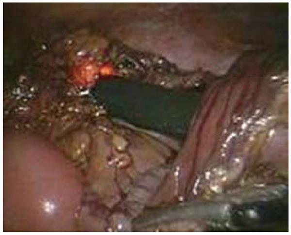
Figure 2 Resected specimen was extracted through the opened colectomy made at the ascending colon using snare forceps via colonoscopy through the anus.
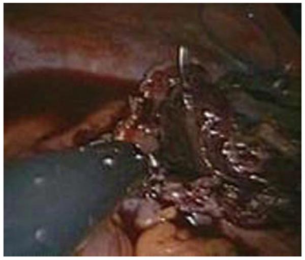
Figure 3 Intracorporeal laparoscopic full thickness V-Loc continuous suturing performed on the oral and anal open ends after side to side anastomosis by a linear stapler.
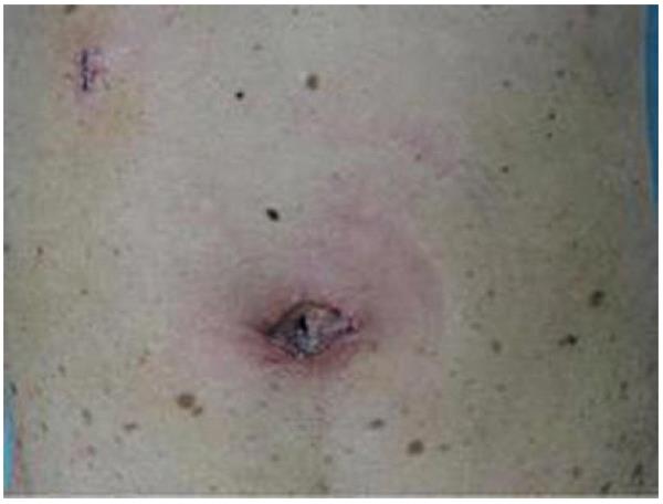
Figure 4 Post operative scars.
- Citation: Takayama S, Hara M, Sato M, Takeyama H. Hybrid natural orifice transluminal endoscopic surgery for ileocecal resection. World J Gastrointest Surg 2012; 4(2): 41-44
- URL: https://www.wjgnet.com/1948-9366/full/v4/i2/41.htm
- DOI: https://dx.doi.org/10.4240/wjgs.v4.i2.41


