Copyright
©2011 Baishideng Publishing Group Co.
World J Gastrointest Surg. Aug 27, 2011; 3(8): 123-127
Published online Aug 27, 2011. doi: 10.4240/wjgs.v3.i8.123
Published online Aug 27, 2011. doi: 10.4240/wjgs.v3.i8.123
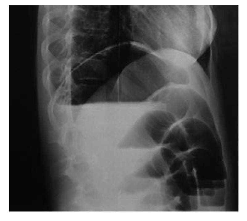
Figure 1 Plain abdominal X-ray demonstrated air fluid levels of the small bowel.
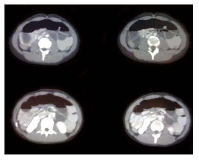
Figure 2 Computed tomography revealed distention of the small intestine at the level of jejunum and ileus.
A mass lesion was identified with concentric rings of fat and soft- tissue attenuation.
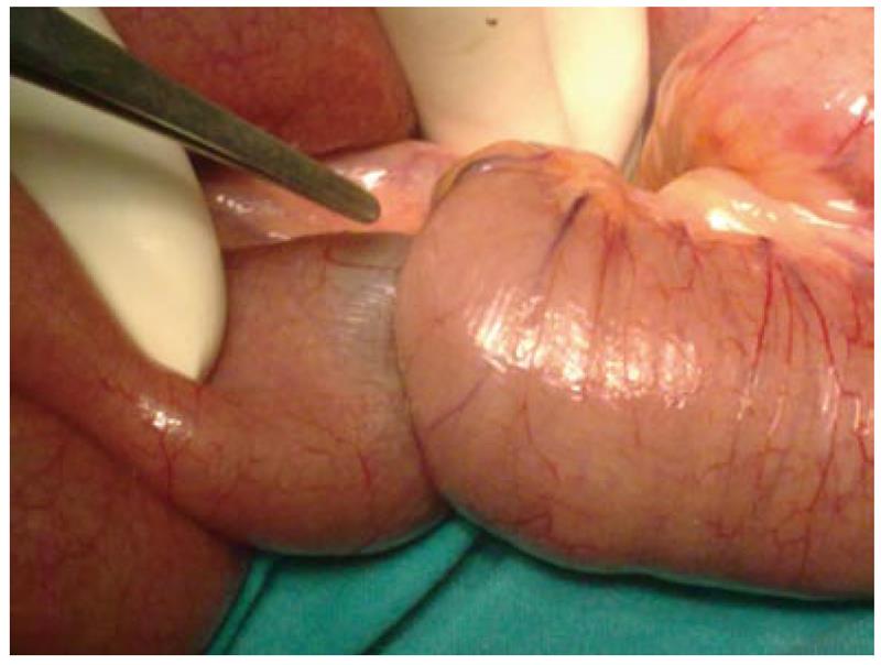
Figure 3 Intussuscepted portion of ileus attributed to inverted Meckel’s diverticulum.
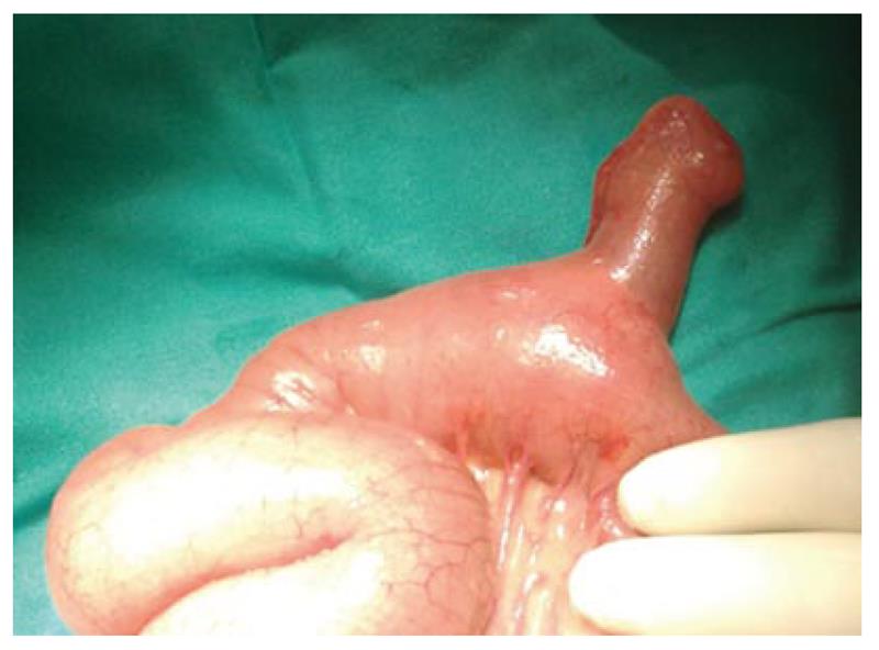
Figure 4 Intraoperative image of a free diverticulum located at an approximate distance of 70 cm from ileocecal valve.
- Citation: Sioka E, Christodoulidis G, Garoufalis G, Zacharoulis D. Inverted Meckel’s diverticulum manifested as adult intussusception: Age does not matter. World J Gastrointest Surg 2011; 3(8): 123-127
- URL: https://www.wjgnet.com/1948-9366/full/v3/i8/123.htm
- DOI: https://dx.doi.org/10.4240/wjgs.v3.i8.123


