Copyright
©2011 Baishideng Publishing Group Co.
World J Gastrointest Surg. Jul 27, 2011; 3(7): 106-109
Published online Jul 27, 2011. doi: 10.4240/wjgs.v3.i7.106
Published online Jul 27, 2011. doi: 10.4240/wjgs.v3.i7.106
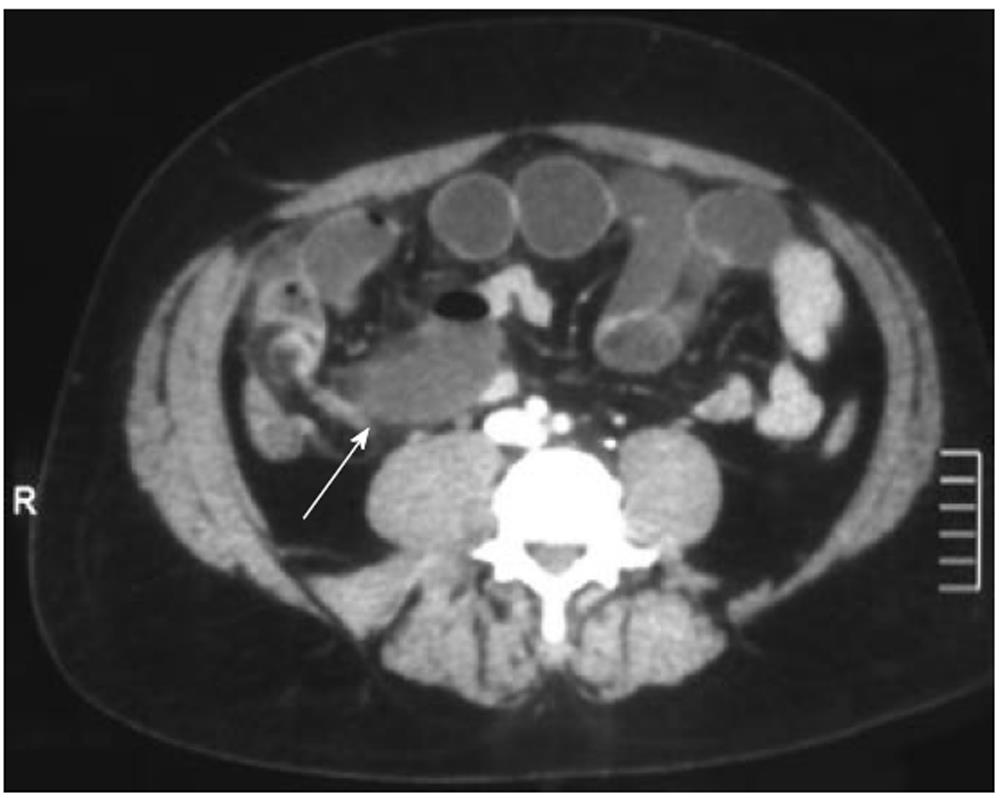
Figure 1 Contrast-enhanced computed tomography images show blind-ending fluid-filled structure (arrow) resulting in small-bowel obstruction.
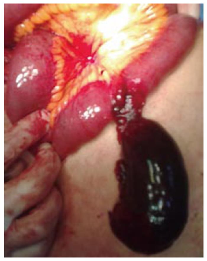
Operative findings confirmed Meckel’s diverticulum.
Figure 2 Segment of ileum with a giant gangrenous Meckel’s diverticulum after derotation.
- Citation: Cartanese C, Petitti T, Marinelli E, Pignatelli A, Martignetti D, Zuccarino M, Ferrozzi L. Intestinal obstruction caused by torsed gangrenous Meckel’s diverticulum encircling terminal ileum. World J Gastrointest Surg 2011; 3(7): 106-109
- URL: https://www.wjgnet.com/1948-9366/full/v3/i7/106.htm
- DOI: https://dx.doi.org/10.4240/wjgs.v3.i7.106


